35 Label The Photomicrograph Of Compact Bone.
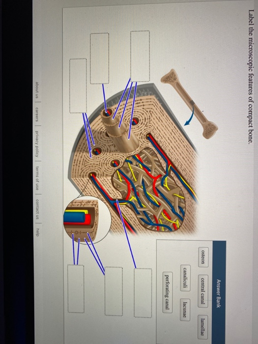
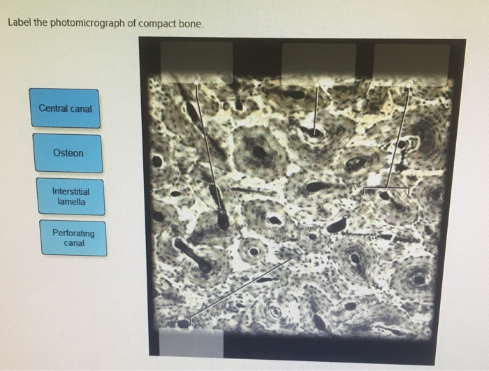
Compact bone forms the surface of all bones Central canal E
This problem has been solved! See the answer
Label the photomicrograph of compact bone.
Contains blood and Periosteum Structure at 1
Label the photomicrograph of compact bone.. On the photomicrograph of bone on the right (365x), identify all structures maned in the key and bracket an osteon Expert Answer Learn vocabulary, terms, and more with flashcards, games, and other study tools
Structure at 4 Structure at 10 Contains blood and
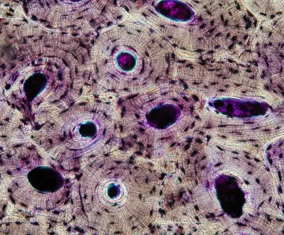
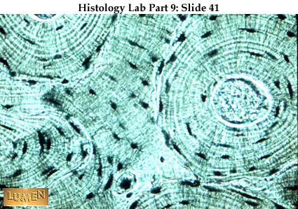
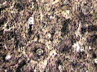
In three dimensions an osteon is cylindrical in shape Osteocyte On each, label the chondrocytes in lacunae and the matrix Blood Vessels
Label The Photomicrograph Of Compact Bone Central Chegg Com
Nerve

Chip Basemap
made of compact bone 6
Answered I Need Help To Label The Structures Bartleby

34 Label The Photomicrograph Of Compact Bone Labels Design

Exercise 8 Cross Section Of Ground Compact Bone Flashcards
describe the histology and location of compact bone and be able to label the following diagram-Haversian system ( osteon ) -Osteocyte-Haversian ( central )canal -Lacunae-Lamellae ( concentric ) -Volkmann's 2

Biology Of Bone Tissue Structure Function And Factors That
Terms in this set (20) Endosteum

Tas Diagram Of Basaltic Samples From The Rift Phases Of The
Haversian Canal

Prepared By Dr Haneen Nur Ppt Download
Perforating Canal
Sedimentary Characteristics Of Microbialites Influenced By
Structure at 6
Untitled
Bone Tissue And Cells Under The Microscope
Osteon

1 Anatomy Lab Skeletal System Histology Of Compact Bone
Structure at 7
Histology Slides 1
Compact Bone

From Buonocore S Pioneering Acid Etch Technique To Self
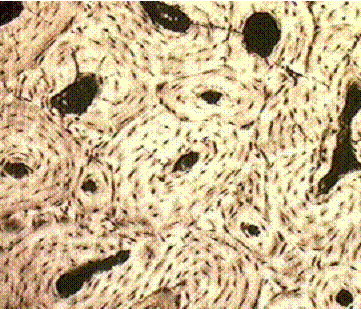
circular channel running longitudinally in the center of an osteon of mature compact bone
Bone Diagenesis Of Tetrapods From The Middle Triassic
Structure at 2

Compact Bone Labeling Quiz
Structure at 3

Estrogen Induces Axonal Outgrowth In The Nucleus
1) Osteon Osteon is the units in the compact tissue

6 3 Bone Structure Anatomy Amp Physiology
Perforating Canal Interstitial Lamella Central Canal Osteon GMA Netrare
Overview Of The Skeleton Classification And Structure Of
A
Bone Compact Ground C S
Microscope at 400X: The outlined area is a cross section of an osteon of compact bone

Tissue Section Of Human Compact Bone Seen Under A Microscope

Compact Bone Microscopic Labeling Diagram Quizlet
Sc 2115 Anatomy And Physiology I
rings of calcified bone matrix surrounding the Haversian canals of compact bone

Respiratory System Sciencedirect
Show transcribed image text
Taphonomy Of Marine Vertebrates Of The Pisco Formation
The bone of the shaft of a long bone is a thick layer of compact bone
Improvement Of Osseointegration By Ultraviolet And Or

Molecular And Morphological Signatures Of Chordate
Periosteum

Compact Bone Lm Stock Image C037 2406 Science Photo
It makes up the outer cortex of all bones and is in immediate contact with the periosteum
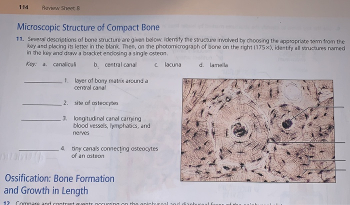
114 Review Sheet 8 Microscopic Structure Of Compact Chegg Com
In long bones, as you move from the outer cortical compact bone to the inner medullary cavity, the bone transitions to spongy bone

A C Ultrastructure Of Bone A Light Microscope Image Of A

Jaypeedigital Ebook Reader
Osteons are cylindrical structures that contain a mineral matrix and living osteocytes connected by canaliculi, which transport blood
Solved Lab 5 Exercise 5 13 In The Photomicrograph Below Of
Identify the two types of cartilage diagrammed below
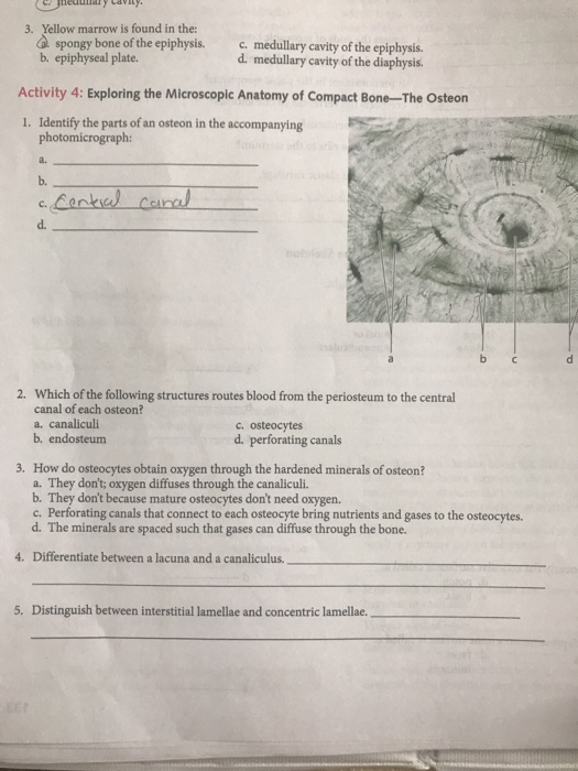
Lacunae D

A Amp P Lab Test 2 Flashcards Quizlet
(a) (b) Start studying 1

Chapter 6 Page 5 Histologyolm 4 0
Slide: Bone, ground, c
0 Response to "35 Label The Photomicrograph Of Compact Bone."
Post a Comment