36 Correctly Label The Following Meninges Of The Brain.
The spinal meninges surround the spinal cord (Figure 13.1a) and are continuous with the cranial meninges, which encircle the brain C. Epidural space - a space between the dura mater and the wall of the vertebral canal Dura mater - The most superficial of the tree spinal meninges is a thick strong layer composed of dense irregular connective tissue. Answer to correctly label the following functional regions of the cerebral cortex. Composed of 3 types of functional areas. There is a visual cortex in each hemisphere of the brain. The majority of the cortex is composed of this area. Saved help save exit submit 4 correctly label the following functional regions of the cerebral cortex.
Label the components of the limbic system in this illustration of a midsagittal section of the brain. Label the selected nerves in the image. Note that not all of the cranial nerves have labels associated with them.. Correctly label the following meninges and associated structures. Correctly label the following figure representing the...

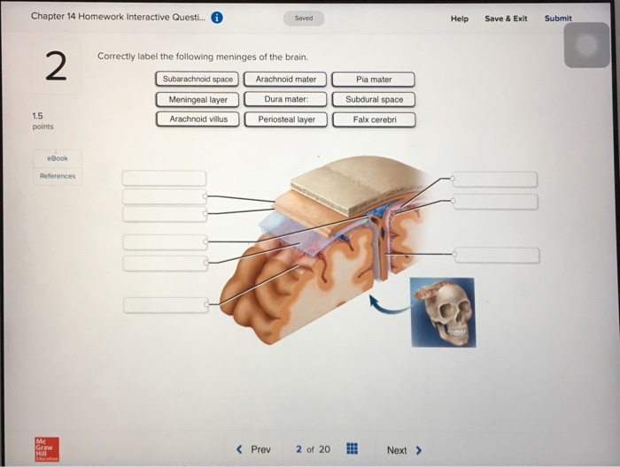
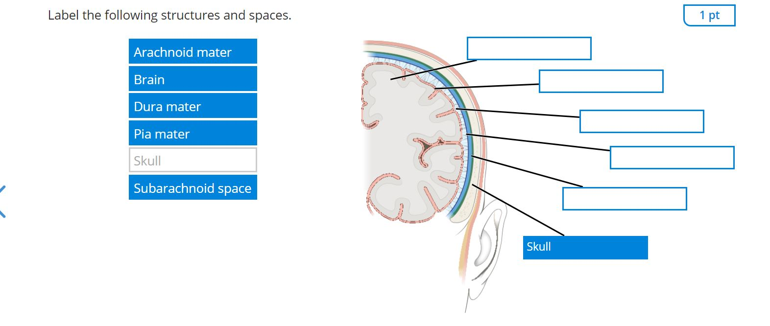
Correctly label the following meninges of the brain.
surrounding the brain and cord (and central canal of the cord) arachnoid Villi ontaining venous blood 10. Label correctly the structures involved with circulation of cerebrospinal fluid on the accompanying diagram. (These struc- tures are identified by leader lines.) V E/JT/QI Cc3QE43íQ/ét- 'V C Correctly label the following functional regions of the cerebral cortex. O o help save exit submt a m correctly label the following structures in the sympathetic nervous system. Two long ganglionated nerve strands one on each side of the vertebral column extending from the base of the skull to the coccyx. Examining the external sheep brain. The tough outer covering of the sheep brain is the dura mater, the outermost meninges membrane covering the brain.Remove the dura mater to see most of the structures of the brain, but remove it carefully, so as to leave all the other structures beneath it intact. Removing the dura mater from the cerebellum at the back of the brain can be tricky.
Correctly label the following meninges of the brain.. ANP1040 Exam 4. Correctly label the following anatomical features of a neuron. Correctly label the structures, areas, and concentrations associated with a cell's electrical charge difference across its membrane. ___ division carries signals to the smooth muscle in the large intestine. Art-labeling Activities. This activity contains 3 questions. Label the regions on the diencephalon and brain stem (posterior view). For each item below, use the pull-down menu to select the letter that labels the correct part of the image. Match the following labels to the proper locations on the sagittal section of the brain. Superior colliculi of the tectal plate The Meninges of the Brain Correctly label the following meninges and associated structures. The Flow of Cerebrospinal Fluid Place a single word into each sentence to make it correct. Not all terms will be used. BI 335 - Advanced Human Anatomy and Physiology Western Oregon University Figure 4: Mid-sagittal section of brain showing diencephalon (includes corpus callosum, fornix, and anterior commissure) Marieb & Hoehn (Human Anatomy and Physiology, 9th ed.) - Figure 12.10 Exercise 2: Utilize the model of the human brain to locate the following structures / landmarks for the
Correctly label the following meninges of the brain. Answer to correctly label the following lymphatics of the thoracic cavity. There are neurolemmocytes or oligodendrocytes at a node of ranvier. Some neurons are specialized to detect stimuli whereas neurons send signals to the effectors of the nervous system. As the lungs expand while. Correctly label the following functional regions of the cerebral cortex. O o help save exit submt a m correctly label the following structures in the sympathetic nervous system. Two long ganglionated nerve strands one on each side of the vertebral column extending from the base of the skull to the coccyx. Anatomy and Physiology questions and answers. Correctly label the following meninges of the brain. Arachnoid villus Arachnoid mater Subdural space Meningeal layer Pia mater Periosteal layer Dura mater: Subarachnoid space Falx cerebri Dura mater: Reset Zoom. Question: Correctly label the following meninges of the brain. Correctly label the following meninges of the brain. Damage to the ___ may affect near vision accommodation.. Consider a situation where a stroke or mechanical trauma has occurred resulting in damage to one of the areas of the brain indicated in the image. Drag each label into the proper location in order to identify the area that would most...
surrounding the brain and cord (and central canal of the cord) arachnoid Villi ontaining venous blood 10. Label correctly the structures involved with circulation of cerebrospinal fluid on the accompanying diagram. (These struc- tures are identified by leader lines.) V E/JT/QI Cc3QE43íQ/ét- 'V C Unformatted text preview: 30. Award: 10.00 points Problems? Adjust credit for all students. Correctly label the following anatomical features of the spinal cord. sheath) (dural mater Dura space Subarachnoid ligaments Denticulate Pia mater Arachnoid cord Spinal Meninges Posterior root ganglion Dura mater (dural sheath) Spinal cord Arachnoid mater Pia mater Fat in epidural space Denticulate. Circulating within the meninges is a liquid substance known as the cerebrospinal fluid (CSF). This fluid cushions the brain and spinal cord to protect them from shocks that could lead to damage. Examining the external sheep brain. The tough outer covering of the sheep brain is the dura mater, the outermost meninges membrane covering the brain.Remove the dura mater to see most of the structures of the brain, but remove it carefully, so as to leave all the other structures beneath it intact. Removing the dura mater from the cerebellum at the back of the brain can be tricky.
Correctly label the following anatomical features of the spinal cord. Drag each label into the appropriate category to identify from which plexus the given nerve emerges. Anatomy and physiology questions and answers. 2a sensory neuron conducts action potentials through the nerve to the spinal cord 3the sensory neuron synapses with an.
The human brain controls nearly every aspect of the human body ranging from physiological functions to cognitive abilities. It functions by receiving and sending signals via neurons to different parts of the body. T he human brain, just like most other mammals, has the same basic structure, but it is better developed than any other mammalian brain.
View the full answer Transcribed image text Drag each label to the appropriate region of the spinal cord Cervical enlargement Lumbar spinal nerves Sacral spinal nerves Lumbosacral enlargement Dural sheath Cervical spinal nerves Terminal Spinal Cord Cross Section The spinal cord which consists of the major nerve tract of vertebrates runs down.
Lobes are simply broad regions of the brain. The cerebrum or brain can be divided into pairs of frontal, temporal, parietal and occipital lobes. Each hemisphere has a frontal, temporal, parietal and occipital lobe. Each lobe may be divided, once again, into areas that serve very specific functions.
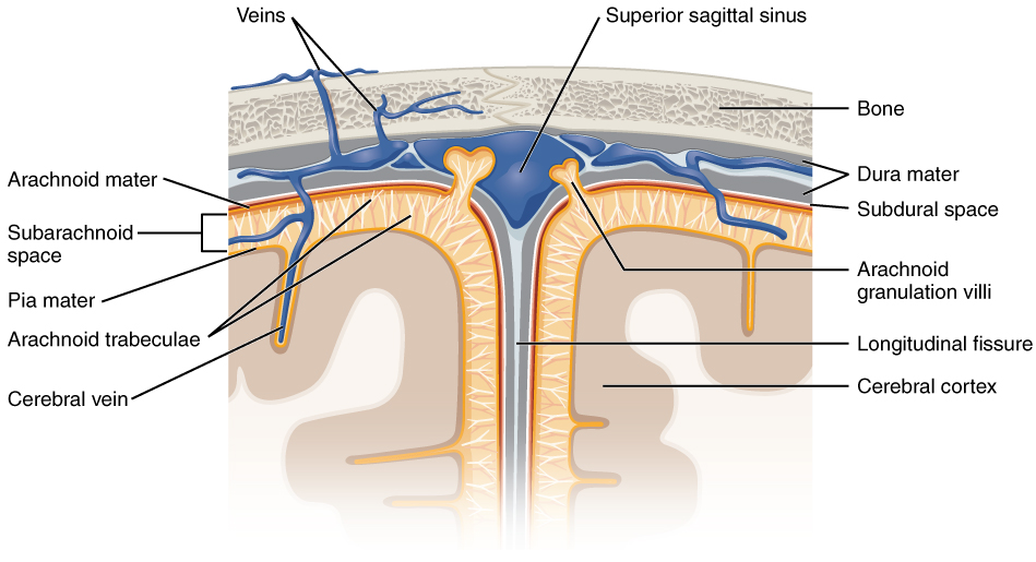
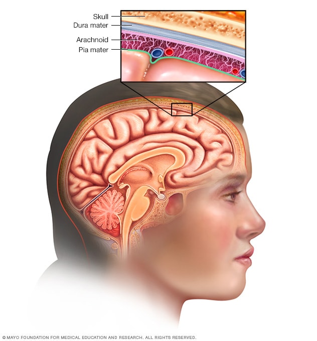
The meninges is a layered unit of membranous connective tissue that covers the brain and spinal cord.These coverings encase central nervous system structures so that they are not in direct contact with the bones of the spinal column or skull. The meninges are composed of three membrane layers known as the dura mater, arachnoid mater, and pia mater.
Correctly label the following meninges of the brain. They protect the brain by housing a fluid filled space and they function as a framework for blood vessels. The arachnoid mater is attached to the pia mater by arachnoid trabeculae which is. Circle and label the following functional areas of the cerebral cortex on brain diagrams.
From outermost to innermost, Meninges around the brain has three layers : 1. The dura mater 2. the arachnoid mater and 3. the pia mater. The dura mater has two layers : periosteal and meningeal. There is a space between the dura mater and the arachnoid mater, called subdural space.
Correctly label the following meninges of the brain. Drag each cranial nerve function to the name of the cranial nerve it corresponds to. Drag each of the following labels into the appropriate box to identify which motor division of the autonomic nervous system is described.
Meninges of the Brain 13. Identify the meningeal (or associated) structures described below: 1. outermost meninx covering the brain; composed of tough fibrous connective tissue 2. innermost meninx covering the brain; delicate and highly vascular 3. structures instrumental in returning cerebrospinal fluid to the venous blood in the dural sinuses
Correctly label the following meninges of the brain.. Consider a situation where a stroke or mechanical trauma has occurred resulting in damage to one of the areas of the brain indicated in the image. Drag each label into the proper location in order to identify the area that would most likely have been affected.
The ventricular system is a set of communicating cavities within the brain. These structures are responsible for the production, transport and removal of cerebrospinal fluid, which bathes the central nervous system. In this article, we shall look at the functions and production of cerebrospinal fluid, and the anatomy of the ventricles that contains it.
Correctly label the following functional regions of the cerebral cortex. Consider a situation where a stroke or mechanical trauma has occurred resulting in damage to one of the areas of the brain indicated in the image. Drag each label into the proper location in order to identify the area that would most likely have been affected.
The Central Nervous System. The CNS consists of the brain and spinal cord, which are located in the dorsal body cavity.The brain is surrounded by the cranium, and the spinal cord is protected by the vertebrae.The brain is continuous with the spinal cord at the foramen magnum. In addition to bone, the CNS is surrounded by connective tissue membranes, called meninges, and by cerebrospinal fluid.
Q&A 2: Meninges, Brain, and Cranial Nerves Overview. STUDY. Flashcards. Learn. Write. Spell. Test. PLAY. Match. Gravity. Created by. daynajc. This quiz includes the following starting on page 25: Meninges Overview, Brain Overview, and Cranial Nerves Overview. Terms in this set (32) What are the meninges? (singular: meninx)
The following topics will be covered: 1. The neurons and supporting cells of the nervous system. Correctly label a diagram of the human brain National Science Education (NSES) Content Standards for grades 9-12... Meninges - A membrane, especially one of the three membranes enclosing the brain and spinal cord in vertebrates.







:background_color(FFFFFF):format(jpeg)/images/library/9598/meninges-and-arachnoid-granulations_english.jpg)




0 Response to "36 Correctly Label The Following Meninges Of The Brain."
Post a Comment