36 Correctly Label The Pectoral And Brachial Muscles.
Get 11 help now from expert anatomy and physiology tutors. Correctly label the muscles of the leg. Correctly label the muscles of the leg. Correctly label the muscles of the leg. Correctly label the muscles of the leg. Expert answer 100 4 ratings previous question next question transcribed image text from this question. Get more help from chegg. The brachial plexus at the level of the trunks and divisions appears as a "bundle of grapes" lateral to the subclavian artery. The lateral end of the transducer is often rotated slightly cephalad to visualize the brachial plexus in a more short-axis plane (perpendicular to its path).
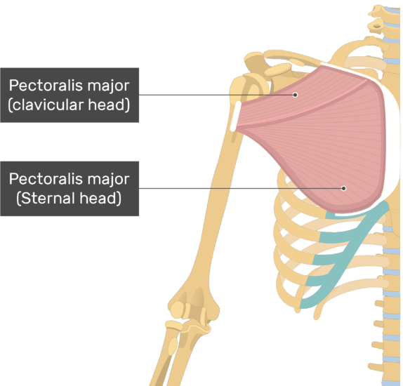
Nerve-supply.-The Pectoralis major is supplied by the lateral and medial pectoral nerves (lateral and medial anterior thoracic nerves); through these it receives filaments from all the nerves entering into the formation of the brachial plexus; the fibers for the clavicular part of the muscle are derived from C. 5 and 6.

Correctly label the pectoral and brachial muscles.
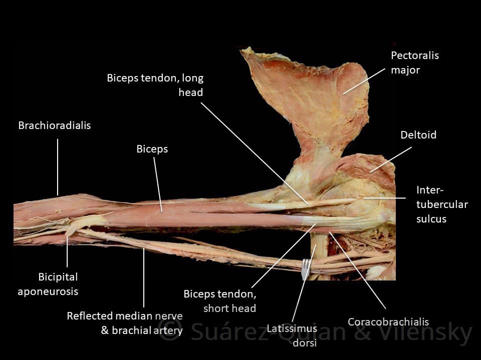
Becomes brachial artery at lower border of teres major Divided into three parts by overlying pectoralis minor First portion, above muscle-gives rise to thoracoacromial a. Second portion, behind muscle-gives rise to lateral thoracic a. Third portion, below muscle-gives rise to subscapular a. divides into throcodorsal a. Correctly label the muscles that act on the hip and femur. Most muscles that act on the femur originate on the hip bone. The two principal anterior muscles are the iliacus, which fills most of the broad iliac fossa of the pelvis, and the psoas major, a thick rounded muscle that arises mainly from the lumbar vertebrae. View Homework Help - 17E60840-5AD9-448F-B7D2-E1DB70FD4769.jpeg from BIO 203 at Bunker Hill Community College. Correctly label the following muscles of the anterior view." Superficial | Deep Tibialis
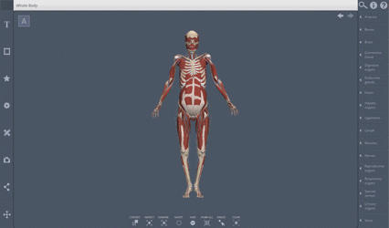
Correctly label the pectoral and brachial muscles.. Unformatted text preview: 37. Award: 1.00 point Problems? Adjust credit for all students. Correctly label the pectoral and brachial muscles. Long head Lateral brachii: Triceps ofscapula Spine major Pectoralis Clavicle Sternum Pectoralis major Supraspinatus Spine of scapula Supraspinatus Sternum Spine of scapula Clavicle Pectoralis major Triceps brachii: Long head Triceps brachii: Lateral head. Upper Arm: Brachial Artery. The brachial artery is a continuation of the axillary artery past the lower border of the teres major. It is the main supply of blood for the arm. Immediately distal to the teres major, the brachial artery gives rise to the profunda brachii (deep artery), which travels with the radial nerve in the radial groove of the humerus and supplies structures in the posterior. We review their content and use your feedback to keep the quality high. Subscapularis: This muscle is a present at the front of shoulder and is involved in the rotation of cuff muscles. This muscle is considered to be the strongest and large.. View the full answer. Transcribed image text: Correctly label the pectoral and brachial muscles. Skeletal Muscles and Body Movement To move the skeleton a skeletal muscle must be attached to a fixed part of the skeleton. The movable end of the muscle that attaches to the bone being pulled is called the muscle’s insertion, and the end of the muscle attached to a fixed (stabilized) bone is called the origin.
Correctly label the pectoral and brachial muscles Select the correct label and characteristic for each of the featured muscles. Label the anatomical features of the bone using the hints provided. Anatomy and Physiology questions and answers. Correctly label the pectoral and brachial muscles. Spine of scapula Triceps brachii Long head Sternum Lateral head Clavicle Pectoralis major Supraspinatus. Question: Correctly label the pectoral and brachial muscles. The medial pectoral nerves arise from the medial cord of the brachial plexus, containing fibers of C8 and T1 spinal nerves. This nerve provides motor innervation to the pectoralis minor muscle and the lower sternocostal part of the pectoralis major muscle. Medial brachial cutaneous nerve The medial brachial cutaneous nerve, also called the. Brachial plexus injury (also known as brachial palsy in. placebo-controlled randomized trial and open-label study to determine whether botulinum toxin type B (BTX-B) is effective in controlling upper-limb spasticity. The study included subjects with an Ashworth Scale score of 2 or more at the elbow, wrist, and fingers.... The muscles ...
Correctly label the pectoral and brachial muscles. Biceps brachii: Short head Long Biceps brachii: Long head Subscapularis Coracobrachialis Brachialis Short head Biceps brachii: Long head Short head Subscapularis Coracobrachialis Brachialis Zoom Explanation: Nine muscles cross the shoulder joint and insert on the humerus. Anatomy and Physiology questions and answers. Correctly label the pectoral and brachial muscles. Brachioradialis Humerus Infraspinatus Anterior Posterior Galater tubercle of humerus Corcobrachialis Teres minor Brachialis Teres major Deltoid Latissimus dorsi. Question: Correctly label the pectoral and brachial muscles. the humerus, deltoid and teres minor muscle. > As the axillary artery emerges from the axilla into the arm it is now called the brachial artery. > brachial artery runs down medial humerus to supply blood to anterior flexor muscles. > deep brachial artery branches off to supply blood to the triceps. At the elbow, Unformatted text preview: 2. Award: 1.00 point Problems? Adjust credit for all students. Correctly label the following features of muscle and fascia. fat Subcutaneous muscle Individual Skin Subcutaneous fat Skin Fascia Subcutaneous fat Vein Fascia Artery Nerve Femur Vein Fascicles Individual muscle Individual muscle Artery Nerve Femur Fascicles Zoom Explanation: The connective tissue.

Chapter 8 10 Flashcards Quizlet
AIMS01-Muscles of Facial Expression: Abnormal Involuntary Movement Scale - Facial and Oral Movements, Muscles of facial expression, e.g., movements of forehead, eyebrows, periorbital area, cheeks. Include frowning, blinking, grimacing of upper face. AIMS - Muscles of Facial Expression: AIMS0102: C102035: AIMS01-Lips and Perioral Area
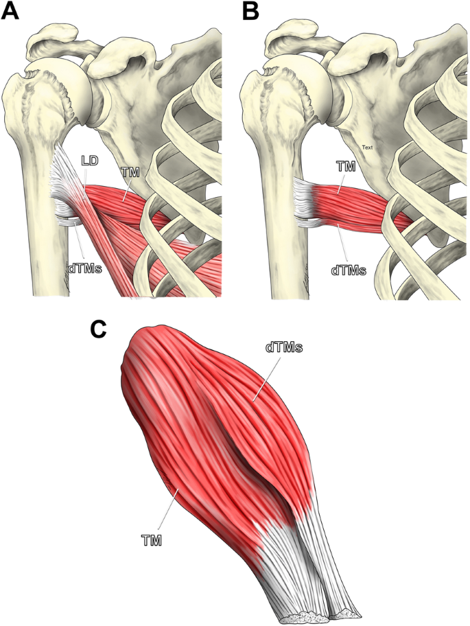
Anatomical Study Of The Teres Major Muscle Description Of An
Correctly label the muscles that act on the hip and femur. Most muscles that act on the femur originate on the hip bone. The two principal anterior muscles are the iliacus, which fills most of the broad iliac fossa of the pelvis, and the psoas major, a thick rounded muscle that arises mainly from the lumbar vertebrae.

Biceps Brachii Attachments Action Amp Innervation
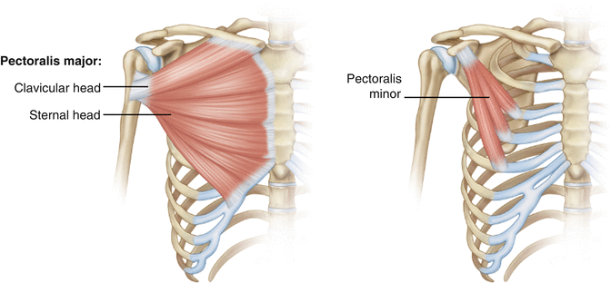
The pectoral region is located on the anterior chest wall. It contains four muscles that exert a force on the upper limb: the pectoralis major, pectoralis minor, serratus anterior and subclavius. In this article, we shall look at the anatomy of the muscles of the pectoral region - their attachments, actions and innervation.
Outline Shape Analysis Of Penguin Humeri A Robust Approach
The thoracic wall is made up of five muscles: the external intercostal muscles, internal intercostal muscles, innermost intercostal muscles, subcostalis, and transversus thoracis. These muscles are primarily responsible for changing the volume of the thoracic cavity during respiration. Other muscles that do not make up the thoracic wall, but attach to it include the pectoralis major and minor.
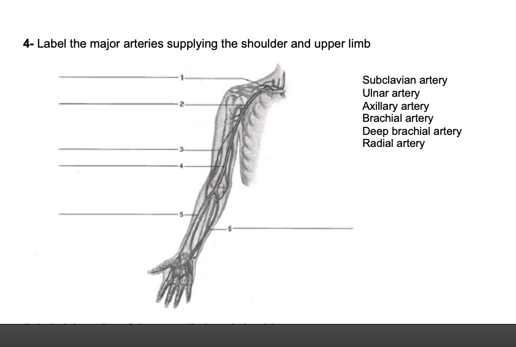
Answered 4 Label The Major Arteries Supplying Bartleby
Clavipectoral fascia is a fibrous sheet present deep to the clavicular part of the pectoralis major muscle. Extent: From clavicle (margins of subclavian groove) above to the axillary fascia below. Muscles enclosed: Subclavius and pectoralis minor. Structures piercing: Lateral pectoral nerve. Cephalic vein.

Neural Crest And The Patterning Of Vertebrate Craniofacial
The primary nerve supply to the pectoralis minor muscle comes via the medial pectoral nerve (C8, T1), one of the minor branches of the brachial plexus that arises from the cervical portion of the spinal cordInnervation to the pectoralis minor is also received from the lateral pectoral nerve, via a communicating branch known as the 'ansa pectoralis', which is usually found anterior to the.
Muscles Of The Upper Arm Biceps Triceps Teachmeanatomy
Unformatted text preview: 36. Award: 1.00 point Problems? Adjust credit for all students. Correctly label the pectoral and brachial muscles. ofhumerus tubercle Greater dorsi Latissimus major minor Teres Deltoid Latissimus dorsi Brachialis Brachioradialis Anterior Deltoid Coracobrachialis Greater tubercle of humerus Infraspinatus Brachialis Posterior Teres minor Greater tubercle of humerus.
30 Correctly Label The Pectoral And Brachial Muscles
Start studying A&P : Assignment 1b. Learn vocabulary, terms, and more with flashcards, games, and other study tools.
Pectoralis Major Muscle Attachment Action Amp Innervation
Start studying Pectoral and Brachial Muscles 5. Learn vocabulary, terms, and more with flashcards, games, and other study tools.

Muscle Anatomy
Academia.edu is a platform for academics to share research papers.

Lecture 2 Thoracic Wall Amp Diaphragm
Jun 03, 2020 · Emory Department of GYNOB on Instagram: “You can’t see it.

Muscle Anatomy
What is the nerve supply of the pectoralis major muscle? medial (C8, T1 - medial cord of brachial plexus) & lateral (C5, C6, C7 - lateral cord of the brachial plexus) pectoral nerves

Muscle Anatomy
Pectoral and Brachial Muscles Actions 1.. Tries. Unlimited Last Played. 27 Jan, 2021 Sound On/Off. From the quiz author. label the actions Remaining 0. Correct 0. Wrong 0. Press play! 0%. 0:00.0. Quit. Again. This game is part of a tournament. You need to be a group member to play the tournament. Join group, and play Just play.

Chapter 8 10 Flashcards Quizlet
The transverse sections below show muscle groups in the upper and lower limbs. The flexors and extensors of the wrist the muscles which flex and extend the fingers of course also move the hand as a whole but in addition to these muscles there are five others two flexor muscles and three extensor muscles which are inserted into the bones of the metacarpus and not into the phalanges.
Deltoid Physiopedia
In the process of doing an axillary lymph node dissection in a 50 year-old patient, the surgery resident cleans the space between the pectoralis major and minor muscles, in an attempt to remove all of the lateral pectoral lymph nodes. Upon recovery it is noted that the patient's lower pectoralis major is paralyzed.
Anatomy And Physiology Lab I On Openalg
Feb 07, 2008 · FUNDAMENTAL OF NURSING PROCEDURE MANUAL for PCL course. Mohd.javed Khan. Akram Khan

32 Correctly Label The Pectoral And Brachial Muscles
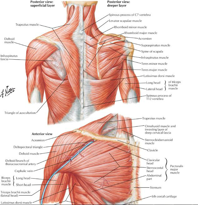
Similar to the muscles that position the pectoral girdle, muscles that cross the shoulder joint and move the humerus bone of the arm include both axial and scapular muscles (Figure 11.4.16 and Figure 11.4.17). The two axial muscles are the pectoralis major and the latissimus dorsi.
Anatomy And Physiology Lab I On Openalg
Correctly label the pectoral and brachial muscles. Show transcribed image text. Correctly label the muscles of the neck back and gluteal region. A large triangular muscle covering the shoulder joint and serving to abduct and flex and extend and rotate the arm. Each of these is a correct listing of muscles attached to the scapula.

Pectoral And Brachial Muscle Actions 3 Quiz
View Homework Help - 17E60840-5AD9-448F-B7D2-E1DB70FD4769.jpeg from BIO 203 at Bunker Hill Community College. Correctly label the following muscles of the anterior view." Superficial | Deep Tibialis

8 2 Bones Of The Upper Limb Anatomy Amp Physiology
Becomes brachial artery at lower border of teres major Divided into three parts by overlying pectoralis minor First portion, above muscle-gives rise to thoracoacromial a. Second portion, behind muscle-gives rise to lateral thoracic a. Third portion, below muscle-gives rise to subscapular a. divides into throcodorsal a.
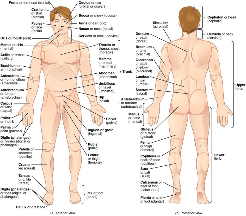
1 4 Anatomical Terminology Fundamentals Of Anatomy And
Anatomy And Physiology Support And Movement The Muscular

Muscles Of The Upper Arm Biceps Triceps Teachmeanatomy
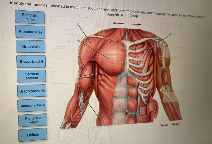
Solved Identify The Muscles Indicated In The Chest Chegg Com
Strength Testing Springerlink
Anatomy Tv Titles

General Anatomy Of The Bull And The Cow Illustrated Atlas

Chapter 8 10 Flashcards Quizlet

Chapter 8 10 Flashcards Quizlet

30 Correctly Label The Following Antagonistic Muscles Of The
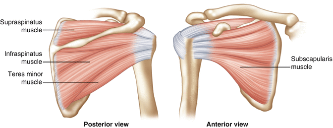
Shoulder Muscles Anatomy And Functions Kenhub

Muscles Of The Pectoral Girdle And Upper Limbs Anatomy And

Pectoralis Major Muscle Wikipedia
Strength Testing Springerlink

Utility Of Ultrasound Guided Injection Of Botulinum Toxin

1 1000 2737 Http Uilis Unsyiah Ac Id Oer Files Original
0 Response to "36 Correctly Label The Pectoral And Brachial Muscles."
Post a Comment