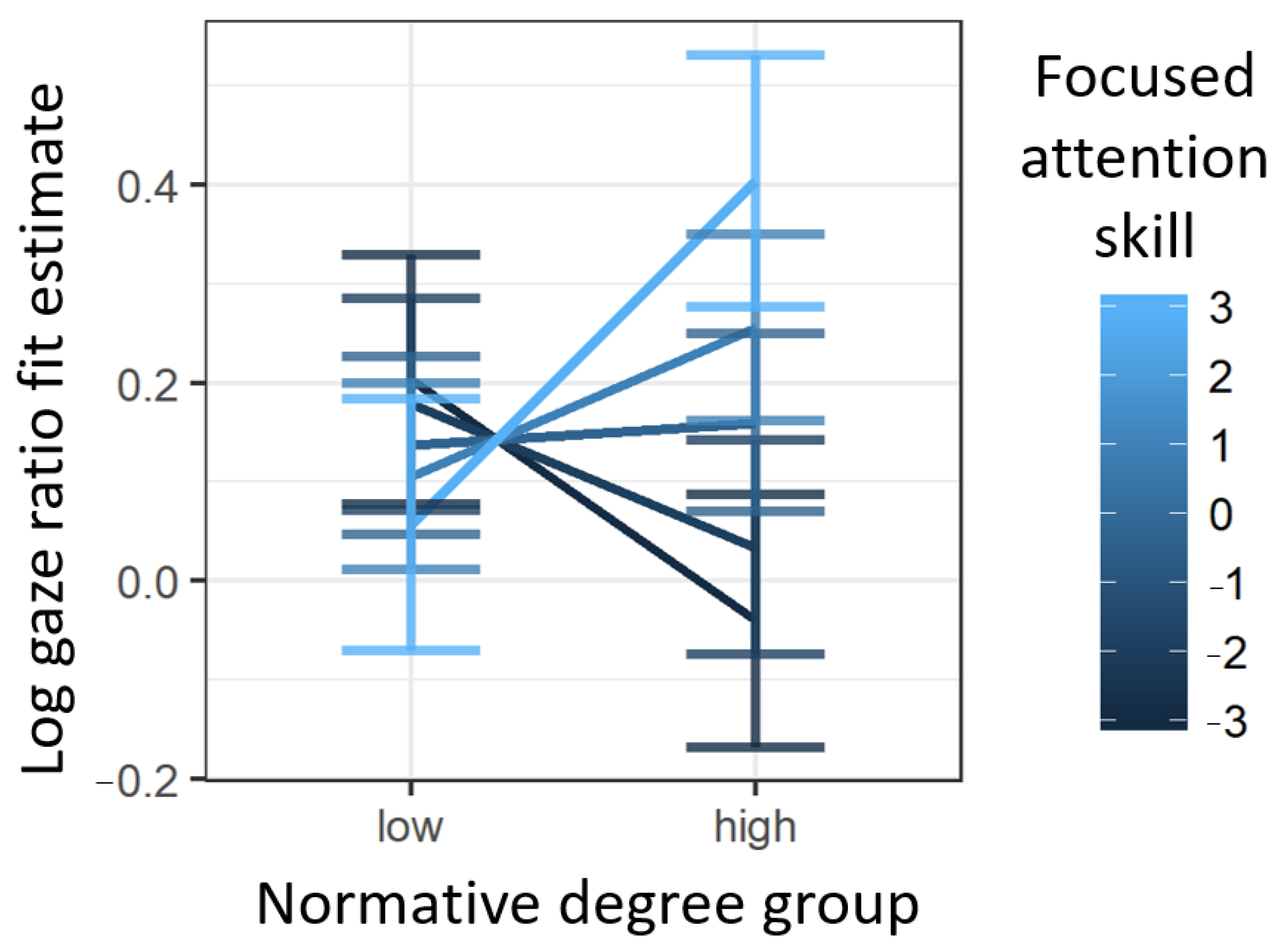
40 Label The Figure With The Items Provided.
QLabel is used for displaying text or an image. No user interaction functionality is provided. The visual appearance of the label can be configured in various ways, and it can be used for specifying a focus mnemonic key for another widget. A QLabel can contain any of the following content types: Content. Setting. The cells in all of the layers except the stratum basale are called keratinocytes. A keratinocyte is a cell that manufactures and stores the protein keratin. Keratin is an intracellular fibrous protein that gives hair, nails, and skin their hardness and water-resistant properties.The keratinocytes in the stratum corneum are dead and regularly slough away, being replaced by cells from the.
Captions for Figures and Tables. In papers written for classes and submitted to journals, every table and figure should include a caption, honoring these common practices: The caption for a figure appears below the graphic; for a table, above. It is easy to get this wrong accidentally. Typically, boldface or underscore the word "Figure" or.
Label the figure with the items provided.
In the figure, angles 4 and 5 are consecutive interior angles. In the figure, angles 5 and 7 are vertical angles. $16:(5 79; Vertical Angle Thm. Cons. Int. s Thm. 5 62/87,21 In the figure, angles 4 and 5 are consecutive interior angles. $16:(5 79; Cons. Int. s Thm. ROADS In the diagram, the guard rail is parallel to Figures should be labeled with a number preceding the table title; tables and figures are numbered independently of one another. Also be sure to include any additional contextual information your viewer needs to understand the figure. For graphs, this may include labels, a legend explaining symbols, and vertical or horizontal tick marks. Figure 2—1 is a diagram of an atom. Select two different colors and use them to color the coding circles and corresponding structures on the figure. Complete this exercise by responding to the questions that follow, referring to the atom in this figure. Insert your answers in the answer blanks provided. O Nucleus Electrons 1. 2. 3. 5. Figure.
Label the figure with the items provided.. across line ℓ. Label the image ˜A′B′′C. Step 2 Translate ˜A′B′C ′ onal g ˝v . Label this image ˜A ″B C . A′ B′ C′ A B C ℓ ˛ν A B C ℓ ˛ν A′ B′ B″ A″ C″ C′ A B ℓ ˛ν If a figure is rotated and then the image is rotated about the same center, a single rotation by the sum of the angles of rotation. Homework i Saved Label the figure with the items provided. Fundus is Uterus is the Fundus is at between the size of a fist the level of the Fundus is ic symphy and resides in xiphoid superior to the and umbilicus the pelvis process of the umbilicus sternum -Diaphragm Xiphold ces process Umbilicus Uterus Pubic - Pubic symphysis symphysis Reset Zoom < Prev 4 of 25 Next > Question: Label the figure with the items provided. Tertiary neurons CN VII CN IX Thalamus Gustatory cortex Secondary neurons Nucleus solitarius. This problem has been solved! See the answer See the answer See the answer done loading. label the figure with the items provided. Figures and graphs usually need to have a label positioned below the figure or graph. As with tables, make sure to number the figures in your document sequentially and to use labels that explain the data clearly and accurately. How to Insert a Label in Microsoft Word. Labels for tables and figures are known as ‘captions’ or ‘legends’.
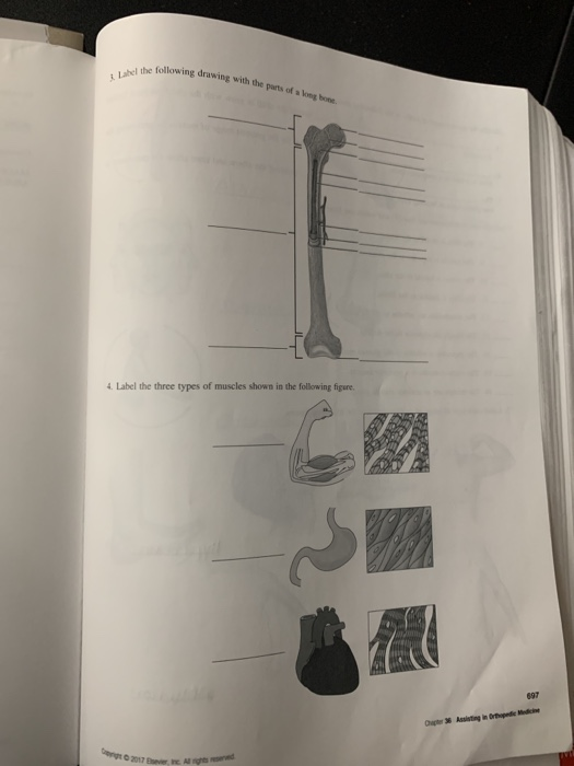
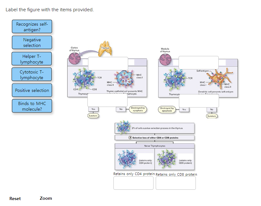
the class label of unknown records. As shown in Figure 4.2, a classification model can be treated as a black box that automatically assigns a class label when presented with the attribute set of an unknown record. Suppose we are given the following characteristics of a creature known as a gila monster: 3. Figure 11—2 is an anterior view of the heart. Identify each numbered structure and write its name in the corresponding numbered space below the figure. Then, select different colors for each structure provided with a color-coding circle, and use them to color the coding circles and corresponding structures on the figure. 10 11 13 O 15 12. Label the figure with the items provided. 1. Helper T-lymphocyte releases IL-2 as an autocrine signal. Label the figure with the items provided. 2. Clones of activated and memory helper T-lymphocytes are produced. Label the figure with the items provided. 3. CD8/TCR binds presented antigen. 1. sponding structures on Figure 6—3. Then bracket and label an A band, an I band, and a sarcomcre, When you have finished, draw a contracted sarcomere in the space beneath the figure and label the same structures, as well as the light and dark bands Z disc Myosin Actin filaments Figure 6—3 1.
direction (Figure 5). Figure 5. Body cavities. Abdominopelvic Quadrants and Regions . The abdominopelvic cavity is quite large and contains many organs, so it is helpful to divide it up into smaller areas for study. The medical scheme divides the abdominal surface (and the abdominopelvic cavity deep to it) into four approximately equal regions Look at Figure B. It shows arteries and veins within the human body. Each artery and vein branches out to tiny capillaries. Write the correct term in each blank to answer the questions or complete the sentence. 1. What pumps blood through your body? _____ 2. Blood vessels that carry blood away from the heart are called _____. 3. a look at the side of your ocular lens and you will notice a label of "10X". This indicates that each ocular lens magnifies the image by a factor of 10 or 10X. OBJECTIVE LENSES - Notice the set of objective lenses on the revolving nosepiece. These lenses allow you to change the degree of magnification. In the figure, angles 4 and 5 are consecutive interior angles. In the figure, angles 5 and 7 are vertical angles. $16:(5 79; Vertical Angle Thm. Cons. Int. s Thm. 5 62/87,21 In the figure, angles 4 and 5 are consecutive interior angles. $16:(5 79; Cons. Int. s Thm. ROADS In the diagram, the guard rail is parallel to
3d4medical From Elsevier On Twitter Complete Anatomy 39 21
Placement of Figures and Tables within the Paper: In manuscripts (e.g. lab papers, drafts), Tables and Figures are usually put on separate pages from text material. In consideration of your readers, place each Table or Figure as near as possible to the place where you first refer to it (e.g., the next page).

Bio 145 Flashcards Quizlet
3.4 Figures and Tables. Visual elements such as graphs, charts, tables, photographs, diagrams, and maps capture your readers' attention and help them to understand your ideas more fully. They are like the illustrations that help tell the story. These visuals help to augment your written ideas and simplify complicated textual descriptions.

Gambar 2 Contoh Analisis Membaca Bersama Mahasiswa Topik
Figure 2—1 is a diagram of an atom. Select two different colors and use them to color the coding circles and corresponding structures on the figure. Complete this exercise by responding to the questions that follow, referring to the atom in this figure. Insert your answers in the answer blanks provided. O Nucleus Electrons 1. 2. 3. 5. Figure.
Solved Label The Figure With The Items Provided Recognizes
5. Figure 6—3 is a diagrammatic representation of a small portion of a relaxed muscle cell (bracket indicates the portion enlarged). First, select different colors for the structures listed below. Use them to color the coding circles and corre- sponding structures on Figure 6—3. Then bracket and label an A band, an I band, and a sarcomere.
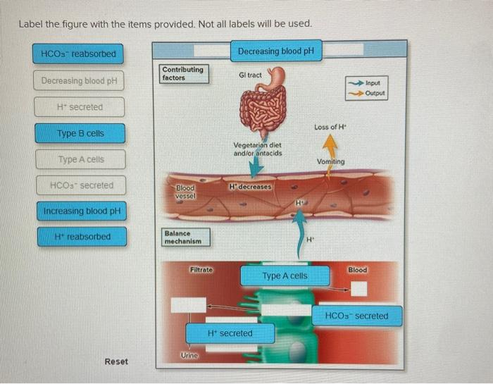
Solved Label The Figure With The Items Provided Not All
An item that may cause confusion when reading electronic prints or schematics is the markings used to show bistable operation. In most cases, bistables will be indicated by
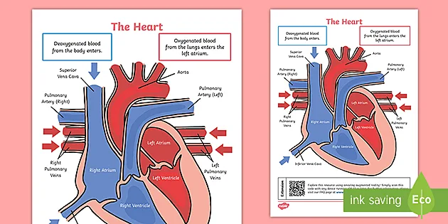
Ks2 The Heart Diagram Qr Labelling Activity
Label the figure with the items provided. Some labels will be used more than once. Label the different types of neuronal pools in the figure. Label the glial cells of the CNS. Identify the functional classification of the neurons in the figure, based on the direction the nerve impulse is traveling relative to the CNS.

Online Shopping Denim And Leather
Label the figure with the items provided Chondrocyte in lacuna begins mitosis Chondrocyte in lacuna begins mitosis. Chondroblasts produce matrix at periphery. Cartilage continues to grow internally Two chondroblasts occupy a lacuna Stem cells in perichondrium begin mitosis Chondroblasts become chondrocytes.
Jtaer Free Full Text Portuguese And Spanish Dmos
Figures should be labeled with a number preceding the table title; tables and figures are numbered independently of one another. Also be sure to include any additional contextual information your viewer needs to understand the figure. For graphs, this may include labels, a legend explaining symbols, and vertical or horizontal tick marks.
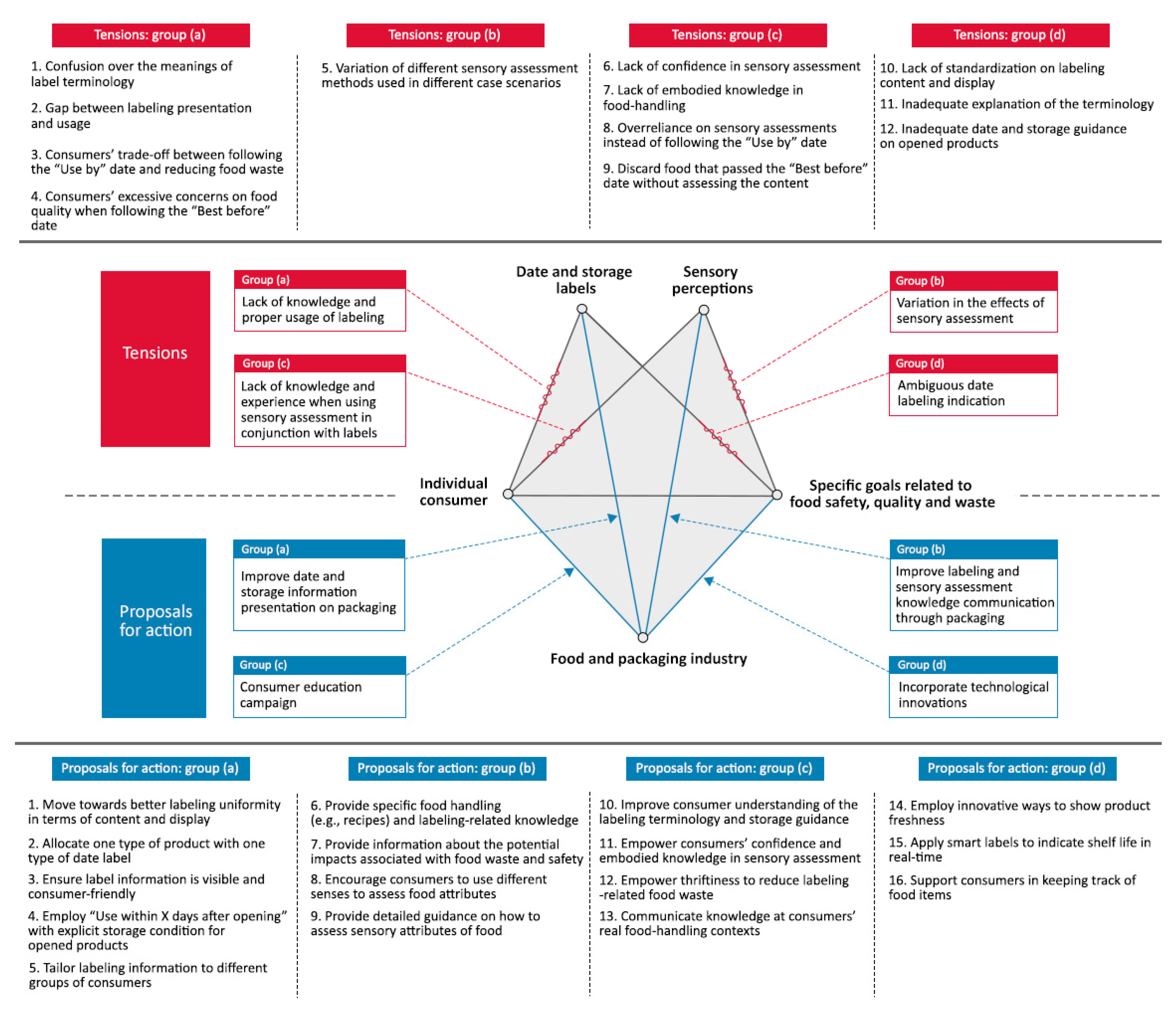
Sustainability Free Full Text Tensions And Opportunities
The following label-reading skills are intended to make it easier for you to use the Nutrition Facts labels to make quick, informed food decisions to help you choose a healthy diet.

Consort Spi 2018 Explanation And Elaboration Guidance For
Label the figure with the items provided. (a) Interstitial Growth (b) Appositional Growth Chondrocyte in lacuna begins mitosis. 0 wenchy Stem cells in perichondrium begin mitosis. Perichondrium Lacus - Chondrocyte New cartilage Chondroblasts become chondrocytes. Diding Metod stem cell Chondrocytes continue producting matrix at periphery.

Chapter 7 Study Set Flashcards Quizlet
Labeling, ranking, sorting, or sentence completion questions. All of these question types require you to position items into an area of the answer box. Answer these kinds of questions on a computer, not on a smartphone. Press Tab to move forward or Shift/Tab to move backwards through the provided answer items.
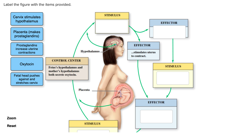
Solved Label The Figure With The Items Provided Stimulus
Figures and graphs usually need to have a label positioned below the figure or graph. Complete each sentence by dragging the proper label into the appropriate position. Answer to label the figure with the items provided. Nutrition Facts Label Basic Requirements Showing Target Items. Solved Label The Figure With The Items Provided 23 To In.

Chapter 12 Question Set Flashcards Quizlet
Label the figure with the items provided. T/F: A normal, healthy cell only displays self-antigens with the MHC class I molecules. True. Alveolar Cells Classify the items with the appropriate cell type. T/F: interferon may induce the degradation of viral RNA or DNA and inhibits the synthesis of viral proteins.
Brain Sciences Free Full Text Perceptual Connectivity
Label the figure with the items provided. cn VII CN IX nucleus, secondary, thalumus, teritary, gustary. A part of the auditory pathway responsible for auditory reflexes is the. not medial.

Aana Journal Course Update For Nurse Anesthetists
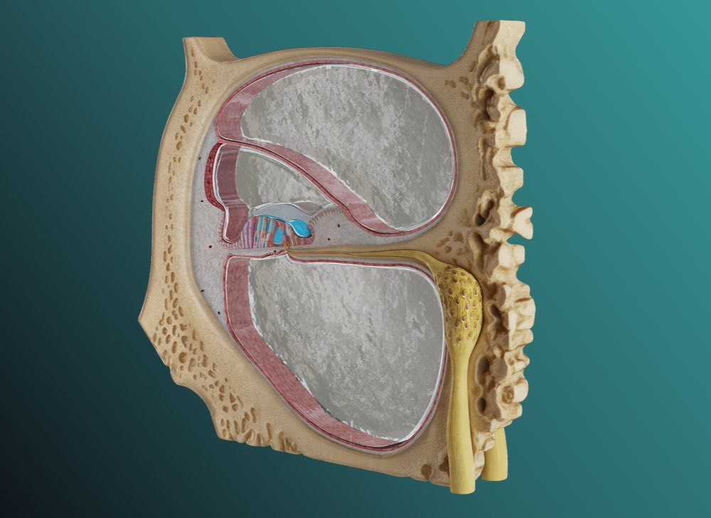
100% (4 ratings) Answer; 1: Compact bone: It is the outer shell of the bone which is hard. 2: Bone resorbed by osteoclast.. View the full answer. Transcribed image text: Label the figure with the items provided. Periosteum Compact bone Bone deposited by osteoblasts Bone resorbed by osteoclasts Medullary cavity. Previous question Next question.
Safe Analysis And Design Of Floor Systems
Figure 2-1: Orthographic projection. 2.2.1) The Six Principle Views The 6 principle views of an orthographic projection are shown in Figure 2-2. Each principle view is created by looking at the object in the directions indicated in Figure 2-2 and drawing what is seen as well as what is hidden from view. Figure 2-2: The six principle views.

Ace Hardware Indonesia Official Store
Label the figure with the items provided. Label the figure with the items provided. Label the molecules on the figure with the terms provided. Label the skeletal system components in the figure with the terms provided. Bone-producing cells are. osteoblasts. Compact bone contains concentric, interstitial, and circumferential.

Taller 1 Ingles Pdf Cell Membrane Osmosis
Anatomy and Physiology questions and answers. Label the figure with the items provided. Colt body Unipolar neuron Dendine Multipolar neuron Anaxonic neuron Bipolar neuron Cell body Dendrites Peripheral process Central process Cell body Single short process Dendrites Cell body Axon Reset Zoom. Question: Label the figure with the items provided.

What S New Matplotlib 3 4 3 Documentation
The cranium (skull) is the skeletal structure of the head that supports the face and protects the brain.It is subdivided into the facial bones and the brain case, or cranial vault (Figure 1).The facial bones underlie the facial structures, form the nasal cavity, enclose the eyeballs, and support the teeth of the upper and lower jaws.

Best Store 2021 1 Type 1 American Silver Eagle Ngc Ms70 Fdi
Draw and label a figure for each relationship. A line in a coordinate plane contains A (0, ±5) and B (3, 1) and a point C that is not collinear with 62/87,21 Plot the points A (0, ±5) and B(3, 1) on a coordinate plane and draw a line through them. Mark the point C anywhere on the coordinate plane except on the.

Chapter 14 Question Set Flashcards Quizlet

Percy Jackson Amp The Olympians Lightning Thief Action Figure W

Camping Cooking Supplies 530ml Gas Stove Tank Oil Containers
Booster Antena Remote Pengganti Boster Anten Putar Merk Intra
Suneducationgroup Com 40cm Halloween Human Skeleton
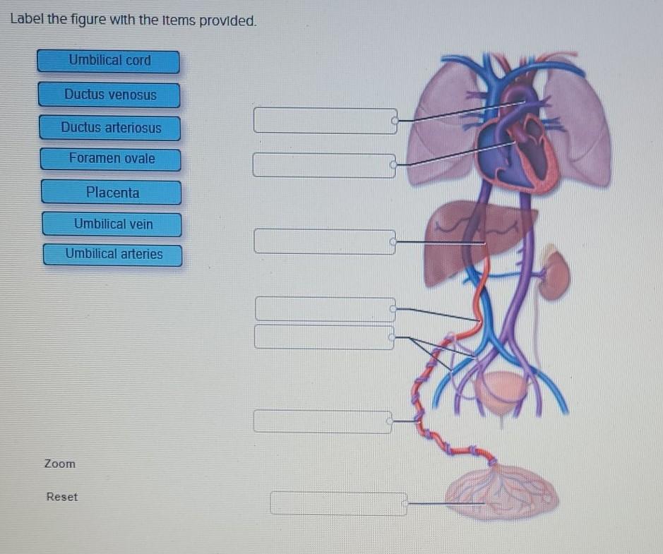
Solved Label The Figure With The Items Provided Umbilical
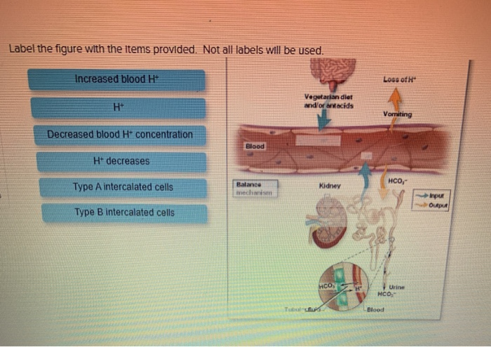
Solved Label The Figure With The Items Provided Not All

Chapter 16 Visual Flashcards Quizlet

Chapter 7 Study Set Flashcards Quizlet

Back Button

Lecture 17 Central Nervous System Cns Bio 004

Safety Tolerability And Immunogenicity Of An Inactivated

Toys Amp Games Tv Amp Movie Character Toys Suneducationgroup Com
32 In The Following Figure Label The Bones Of The Extremities

Three Modeling Applications To Promote Automatic Item
Pfizer Biontech Covid 19 Vaccine
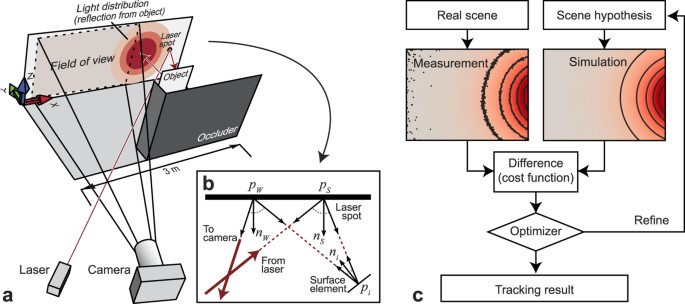
Tracking Objects Outside The Line Of Sight Using 2d Intensity
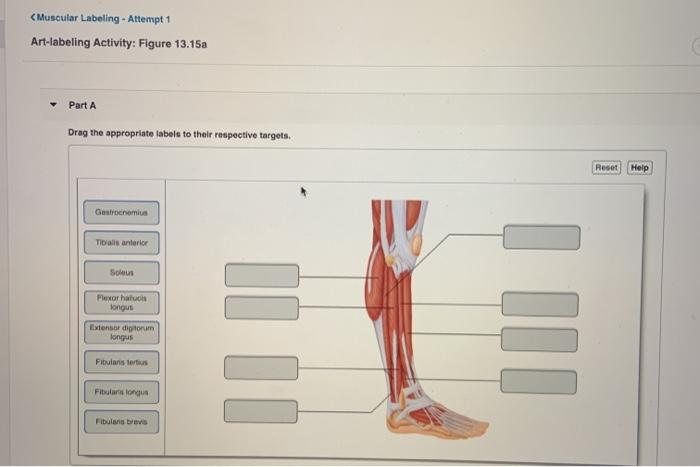
Solved Lt Muscular Labeling Attempt 1 Art Labeling Activity
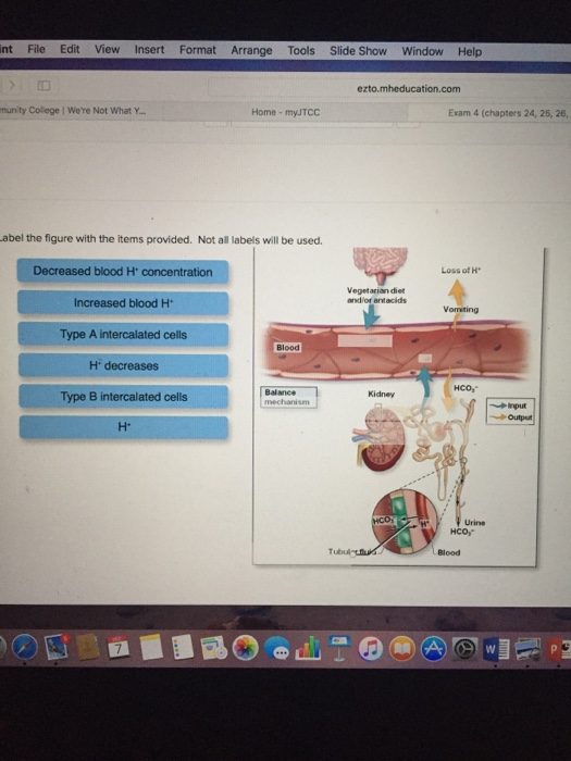
Solved Label The Figure With The Items Provided Not All
Kamus Inggris Indonesia Pdf
0 Response to "40 Label The Figure With The Items Provided."
Post a Comment