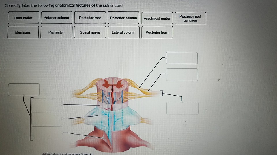
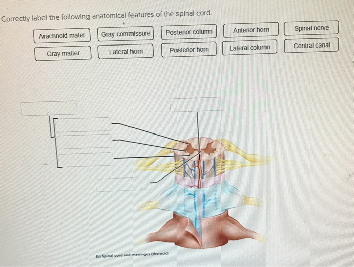
37 correctly label the following anatomical features of the spinal cord.
quizlet.com › 568717950 › chapter-14-question-setChapter 14 Question Set Flashcards - Quizlet Correctly identify the function of each structure that comprises a tendon reflex by dragging the appropriate label into place. Label the structures of the spinal cord. Label the spinal cord meninges and spaces. Label the white and gray matter components in the figure. Label the primary nerves of the lumbar plexus. PDF Chap1-anatomical terminology [Compatibility - LA Mission vertebral or spinal cavity (which holds the spinal cord). b) The ventral cavity: located toward the front of the body, is divided into abdominopelvic cavity and thoracic cavity by the diaphragm. The abdominopelvic cavity is subdivided into abdominal cavity (which holds liver, gallbladder, stomach, pancreas, spleen, kidney,
Anatomy of the Spinal Cord (Section 2, Chapter 3 ... The spinal cord is a cylindrical structure of nervous tissue composed of white and gray matter, is uniformly organized and is divided into four regions: cervical (C), thoracic (T), lumbar (L) and sacral (S), (Figure 3.1), each of which is comprised of several segments.
Correctly label the following anatomical features of the spinal cord.
Spinal Cord - Anatomy, Structure, Function, & Diagram Spinal Cord Anatomy In adults, the spinal cord is usually 40cm long and 2cm wide. It forms a vital link between the brain and the body. The spinal cord is divided into five different parts. Sacral cord Lumbar cord Thoracic cord Cervical cord Coccygeal Several spinal nerves emerge out of each segment of the spinal cord. The spinal cord | Human Anatomy and Physiology Lab (BSB 141) Human Anatomy and Physiology Lab (BSB 141) Module 10: The Nervous System. Search for: The spinal cord. Information. The spinal cord in cross-section has a central region of darker gray matter and the rest is lighter white matter. The gray matter is made up of neuroglia cells and neuron cell bodies. The white matter is made up of neuron axons ... Drag The Correct Label To The Appropriate Location To ... Correctly label the following anatomical features of the thoracic cavity. Label the general pattern of neurons and neurotransmitters associated with the autonomic nervous system. Drag each label into the appropriate position in order to identify whether the structure is an actual part of the digestive tract or an accessory structure.
Correctly label the following anatomical features of the spinal cord.. Chapter 13 Worksheet Flashcards | Quizlet Gravity The spinal cord serves four principle functions: conduction, neural integration, locomotion and reflexes. Click card to see definition 👆 true Click again to see term 👆 1/36 Previous ← Next → Flip Space Sets found in the same folder Chapter 3 Worksheet 50 terms sonjamilosavljevic Module 2 Skeletal System 127 terms quizlette6443568 PLUS › homework-help › questions-andSolved Correctly label the following anatomical features of ... Correctly label the following anatomical features of the spinal cord. 26 Pimate Dura materidura shout Arachnoid mater Meninges Spinal cord Farinebidural space Derttelaments Subdural cu ganglion 1 points Posterior References Meninges Anterior (*) Spinal cord and wertebra (cervical This is the most superficial covering of the spinal cord Reset Zoom. › homework-help › questions-andSolved Correctly label the following anatomical features of ... Anatomy and Physiology Correctly label the following anatomical features of the spinal cord. Spinal nerve Posterior root ganglion Posterior horn Anterior median fissure Lateral hom Anterior horn Central canal Posterior root Posterior median sulcus Spinal nerve Central canal Posterior median sulcus Anterior median fissure Posterior root (1) Spinal cord and meningen (thoracic) Spinal Anatomy | Vertebral Column - SpineUniverse The spinal column (or vertebral column) extends from the skull to the pelvis and is made up of 33 individual bones termed vertebrae. The vertebrae are stacked on top of each other group into four regions: Term. # of Vertebrae. Body Area.
Comments on: Correctly label the following anatomical ... Knowledge for Noob ... Comments on: Correctly label the following anatomical features of the spinal cord. PDF BIO 113 LAB 1. Anatomical Terminology, Positions, Planes ... Surface Anatomy . Body surfaces provide a number of visible landmarks that can be used to study the body. Several of these are described on the following pages. Locating Body Landmarks . Anterior Body Landmarks . Identify and use anatomical terms to correctly label the following regions on Figure 1: Calculate the peak wavelength given off from the following ... Correctly label the following anatomical features of the spinal cord. by soetrust. Post Navigation. Previous Post How many slices is 2 ounces of ham? Next Post how big is 500 square feet? Leave a comment Cancel comment. Your email address will not be published. Required fields are marked * Comment * Spine Structure & Function: Parts & Segments, Spine ... Spinal cord and nerves: The spinal cord is a column of nerves that travels through the spinal canal. The cord extends from the skull to the lower back. Thirty-one pairs of nerves branch out through vertebral openings (the neural foramen). These nerves carry messages between the brain and muscles.
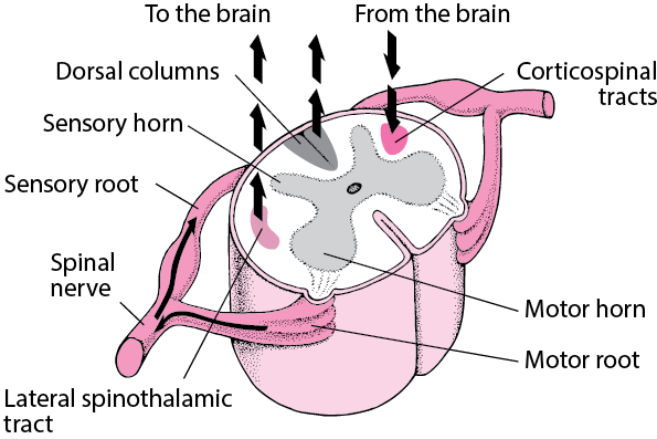
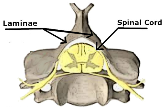
What Are The 5 Sections Of The Spine? Spinal Column Anatomy Spinal Cord & Nerves. The length of the spinal cord is approximately 45 cm in men and 43 cm in women. The diameter ranges from 13 mm in the cervical and lumbar regions to 6.4 mm in the thoracic area. The cord is protected within the spinal canal and runs from the brainstem to the lumbar area where the cord fibres separate. PDF The Nervous System: Spinal Nerves - Napa Valley College Gross Anatomy of the Spinal Cord •Features of the Spinal Cord •Transverse view •White matter •Gray matter •Central canal •Dorsal root and ventral root: merge to form a spinal nerve •Dorsal root is sensory: axons extend from the soma within the dorsal root ganglion •Ventral root is motor fornoob.com › correctly-label-the-followingCorrectly label the following anatomical features of the ... Correctly label the following anatomical features of the spinal cord. Answer Labelled on the left side from top to bottom as 1,2,3 and on right side from top to bottom as 4,5,6,7 1. Posterior horn -it is the sensory horn 2. Gray commisure 3. Anterior median fissure 4. Posterior funiculus - it is the white matter of the spinal cord 5. 14.3 The Brain and Spinal Cord - Anatomy & Physiology The spinal cord is a single structure, whereas the adult brain is described in terms of four major regions: the cerebrum, the diencephalon, the brain stem, and the cerebellum. A person's conscious experiences are based on neural activity in the brain. The regulation of homeostasis is governed by a specialized region in the brain.
Understanding Spinal Anatomy: Regions of the Spine ... Understanding Spinal Anatomy: Regions of the Spine - Cervical, Thoracic, Lumbar, Sacral. The regions of the spine consist of the cervical, thoracic, lumbar, and sacral. Cervical Spine. The neck region of the spine is known as the Cervical Spine. This region consists of seven vertebrae, which are abbreviated C1 through C7 (top to bottom).
AHCDW9Notes30.pdf - 30. Award: 10.00 points Problems ... Correctly label the following anatomical features of the spinal cord. Explanation: The brain and spinal cord are covered by three fibrous membranes that lie between the nervous tissue and bone. These layers and the spaces between them help to protect the delicate nervous tissue from abrasion and other trauma. Collectively, they are called meninges.
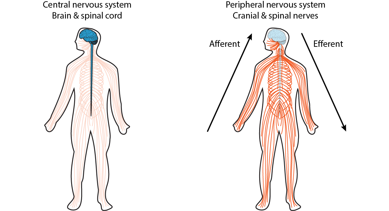
PDF Distance Learning Program Anatomy of the Human Brain/Sheep ... The following topics will be covered: 1. The neurons and supporting cells of the nervous system 2. Organization of the nervous system (the central and peripheral nervous systems) 4. Protective coverings of the brain 5. Brain Anatomy, including cerebral hemispheres, cerebellum and brain stem 6. Spinal Cord Anatomy 7.
Spinal cord: Anatomy, structure, tracts and function | Kenhub The spinal cord is made of gray and white matter just like other parts of the CNS. It shows four surfaces: anterior, posterior, and two lateral. They feature fissures (anterior) and sulci (anterolateral, posterolateral, and posterior). Spinal cord (cross section)
Neuropsychology (PSYC 3311) - East Carolina University Anatomical location Function: III. Drawing and labeling. Draw and label clearly) a brain from a lateral perspective. Label the following structures and anatomical features (8 points): 1) central fissure 2) lateral fissure 3) frontal lobe 4) parietal lobe 5) occipital lobe 6) temporal lobe 7) primary somatosensory cortex 8) primary auditory cortex.
Correctly Label The Following Anatomical Features Of The ... Correctly Label The Following Anatomical Features Of The Spinal Cord. Lateral Funiculus Posterior Root Of Spinal Nerve Posterior Funiculus Posterior Horn Anterior Median Fissure Spinal Nerve Gray Commissure Spinal Nerve (B) Spinal Cord And Meninges (Thoracic) Feb 19 2022 01:49 PM. Solution.pdf.
› file › 30098411AHCDW9Notes34.pdf - 34. Award: 10.00 points Problems? Adjust ... Correctly label the following anatomical features of the spinal cord. Explanation: The spinal cord is wrapped in a threelater protective covering called the meninges. In a crosssectional view, one can also contrast the white matter to the gray matter of the spinal cord. The white matter is arranged in peripherallylocated columns of myelinated nerve
33 Correctly Label The Following Anatomical Features Of A ... Correctly label the following anatomical features of the surface of the brain. Trigger axon hillock trigger zone. 7 this neuron part sends on messages to other neurons. 8 this neuron part gives messages to muscle tissue. Anatomy of a neuron. Correctly label the following anatomical features of a neuron.
soetrust.org › misc › correctly-label-the-followingCorrectly label the following anatomical features of the ... Feb 26, 2022 · Misc. Correctly label the following anatomical features of the spinal cord. by soetrust February 26, 2022 Leave a reply 1. SOMEONE ASKED. Correctly label the following anatomical features of the spinal cord.
PDF Brain Anatomy - Wou BI 335 - Advanced Human Anatomy and Physiology Western Oregon University Figure 4: Mid-sagittal section of brain showing diencephalon (includes corpus callosum, fornix, and anterior commissure) Marieb & Hoehn (Human Anatomy and Physiology, 9th ed.) - Figure 12.10 Exercise 2: Utilize the model of the human brain to locate the following structures / landmarks for the
opilizeb.blogspot.com › 2018/09/30-correctly-label30 Correctly Label The Following Anatomical Features Of The ... Correctly label the following anatomical features of the spinal cord. Correctly identify and label the structures associated with the anatomy of a ganglion. Drag each label into the appropriate category to identify from which plexus the given nerve emerges. 14 1 Sensory Perception Anatomy And Physiology
Free Science Flashcards about ANP1040 Exam 4 Correctly label the following anatomical features of the spinal cord. Posterior root ganglion, Spinal cord, Spinous process of vertebra, Epidural space, Pia mater, Dura mater (dural sheath), Arachnoid mater, Spinal nerve, Vertebral body, Subarachnoid space: Correctly label the following anatomical features of a nerve.
Drag The Correct Label To The Appropriate Location To ... Correctly label the following anatomical features of the thoracic cavity. Label the general pattern of neurons and neurotransmitters associated with the autonomic nervous system. Drag each label into the appropriate position in order to identify whether the structure is an actual part of the digestive tract or an accessory structure.
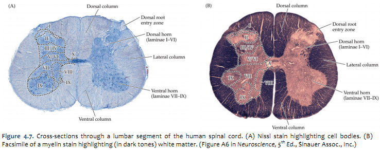
The spinal cord | Human Anatomy and Physiology Lab (BSB 141) Human Anatomy and Physiology Lab (BSB 141) Module 10: The Nervous System. Search for: The spinal cord. Information. The spinal cord in cross-section has a central region of darker gray matter and the rest is lighter white matter. The gray matter is made up of neuroglia cells and neuron cell bodies. The white matter is made up of neuron axons ...
Spinal Cord - Anatomy, Structure, Function, & Diagram Spinal Cord Anatomy In adults, the spinal cord is usually 40cm long and 2cm wide. It forms a vital link between the brain and the body. The spinal cord is divided into five different parts. Sacral cord Lumbar cord Thoracic cord Cervical cord Coccygeal Several spinal nerves emerge out of each segment of the spinal cord.



:background_color(FFFFFF):format(jpeg)/images/library/11473/spinal-membranes-and-nerve-roots_english.jpg)










0 Response to "37 correctly label the following anatomical features of the spinal cord."
Post a Comment