41 correctly label the following anatomical features of the elbow joint.
Chapter 8 Joints Flashcards by Cait Ainger - Brainscape Study Chapter 8 Joints flashcards from Cait Ainger's Georgian College class online, or in Brainscape's iPhone or Android app. Learn faster with spaced repetition. Fibrous Joints - TeachMeAnatomy - Making Anatomy Simple A joint is defined as a connection between two bones in the skeletal system.. Joints can be classified by the type of the tissue present (fibrous, cartilaginous or synovial), or by the degree of movement permitted (synarthrosis, amphiarthrosis or diarthrosis).. In this article, we shall look at the classification of joints in the human body.
Free Science Flashcards about ANP1040 Exam 3 - StudyStack Correctly label the following anatomical parts of a flat bone. Suture, Outer compact bone, Spongy bone, Trabeculae, Inner compact bone: Classify the following images into the types of synovial joints they represent. Saddle, Pivot, Ball & Socket, Hinge, Plane, Condylar: Correctly label the following anatomical features of the elbow joint.
Correctly label the following anatomical features of the elbow joint.
Chart of Major Muscles on the Front of the Body with Labels Anatomical terms allow health care professionals to accurately communicate to others which part of the body may be affected by disorder or a disease. Ultimately, communicating using anatomical terms makes it easy to communicate descriptions of body areas regardless of the individual's position. ... the wrist is distal to the elbow joint ... Solved Correctly label the following anatomical features ... Question: Correctly label the following anatomical features of the elbow joint. Joint capsule Radius Lateral epicondyle Radial collateral ligament Humerus Ulna Anular ligament Medial epicondyle (a) Anterior view This problem has been solved! See the answer Show transcribed image text Expert Answer 100% (8 ratings) › 43265538 › Colby_Lynn_Allen(PDF) Colby Lynn Allen Kisner Carolyn Therapeutic exercise ... Enter the email address you signed up with and we'll email you a reset link.
Correctly label the following anatomical features of the elbow joint.. achieveressays.comAchiever Essays - Your favorite homework help service Our academic writers and editors make the necessary changes to your paper so that it is polished. We also format your document by correctly quoting the sources and creating reference lists in the formats APA, Harvard, MLA, Chicago / Turabian. quizlet.com › 303139357 › joints-homework-flash-cardsJoints Homework Flashcards | Quizlet Label the ligaments and other features shown in this medial view of the elbow. Match each ligament (or pair of ligaments) with the correct joint. Name the ligament that holds the head of the radius in place within the elbow joint. › 40412391 › Clinical_case_studies(PDF) Clinical case studies in physiotherapy - Academia.edu Enter the email address you signed up with and we'll email you a reset link. Drag Each Label to the Location of Each Structure Described Correctly label the following major systemic arteries. Each label can be used more than once. Drag and drop each label identifying the cerebral area that if injured would result in the functional disturbance described. Correctly identify and label the structures associated with the anatomy of a spinal nerve and ganglion.
Anatomy of Selected Synovial Joints | Anatomy and Physiology I (a) The elbow is a hinge joint that allows only for flexion and extension of the forearm. (b) It is supported by the ulnar and radial collateral ligaments. (c) The annular ligament supports the head of the radius at the proximal radioulnar joint, the pivot joint that allows for rotation of the radius. Tooth anatomy: Structure, parts, types and functions | Kenhub Anatomy of the tooth Author: Alexandra Sieroslawska MD • Reviewer: Dimitrios Mytilinaios MD, PhD Last reviewed: October 28, 2021 Reading time: 6 minutes The tooth is one of the most individual and complex anatomical as well as histological structures in the body. The tissue composition of a tooth is only found within the oral cavity and is limited to the dental structures. PDF BIO 113 LAB 1. Anatomical Terminology, Positions, Planes ... Surface Anatomy . Body surfaces provide a number of visible landmarks that can be used to study the body. Several of these are described on the following pages. Locating Body Landmarks . Anterior Body Landmarks . Identify and use anatomical terms to correctly label the following regions on Figure 1: Humerus: Anatomy and clinical notes | Kenhub It consists of a proximal end, a shaft and a distal end, all which contain important anatomical landmarks. The humerus articulates with the scapula proximally at the glenohumeral joint so it participates in the movements of the shoulder . Also, the humerus has distal articulations with the radius and ulna at the elbow joint .
Classification of Joints | Boundless Anatomy and Physiology A joint, also known as an articulation or articular surface, is a connection that occurs between bones in the skeletal system. Joints provide the means for movement. The type and characteristics of a given joint determines its degree and type of movement. Joints can be classified based on structure and function. Chapter 9 QS Anatomy (Joints) Flashcards - Quizlet A gomphosis is functionally classified as a synarthrosis Correctly label the following anatomical features of the elbow joint. ulna, ulnar collateral ligament, anular ligament, radius, articular capsule _____ synovial joints are diarthroses. all The elbow joint is composed of two articulations. True When kicking a ball forward, the knee is extended Correctly label the following anatomical features of the ... The forearm bone is part of the elbow joint. The bicep muscle in the forearm bends it against the weight of the forearm and a weight that the hand is … Chemical messengers that carry important information from one cell to another are called __________. A. Reuptakers B. Neural receptors C. Neurotransmi … 9.4 Synovial Joints - Anatomy & Physiology (a) Pivot joints allow for rotation around an axis, such as between the first and second cervical vertebrae, which allows for side-to-side rotation of the head. (b) The hinge joint of the elbow works like a door hinge. (c) The articulation between the trapezium carpal bone and the first metacarpal bone at the base of the thumb is a saddle joint.
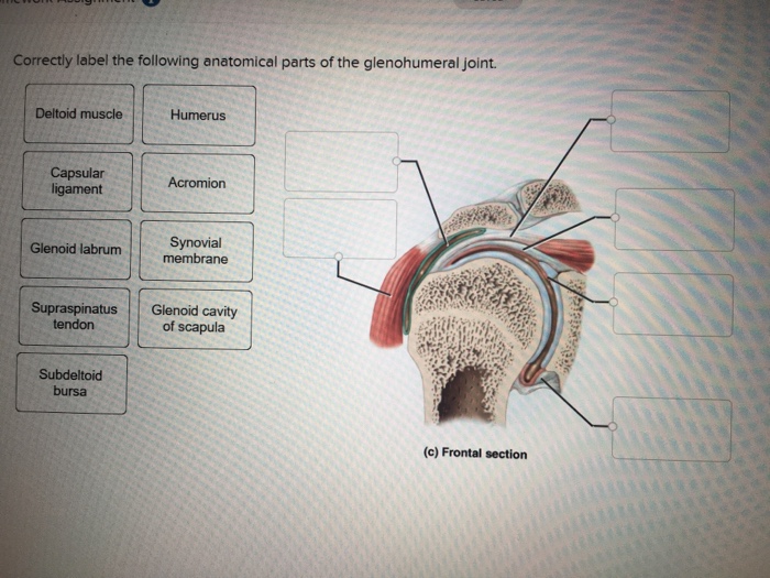
34 Correctly Label The Following Anatomical Parts Of The Glenohumeral Joint. - Labels For Your Ideas
courseworkhero.co.ukCoursework Hero - We provide solutions to students Please Use Our Service If You’re: Wishing for a unique insight into a subject matter for your subsequent individual research; Looking to expand your knowledge on a particular subject matter;
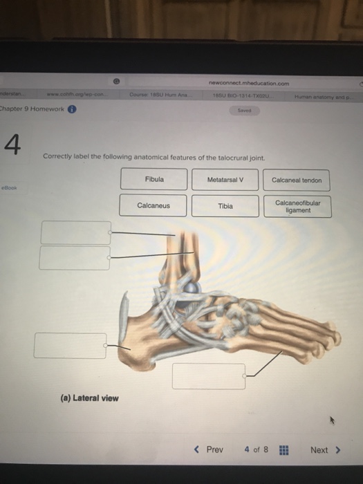
Solved: Newconnect.mheducation.com Course: 18SU Hum Ana SU... | Chegg.com
Knee Joint - Anatomy Pictures and Information The joint-forming surfaces of each bone are covered in a thin layer of hyaline cartilage that gives them an extremely smooth surface and protects the underlying bone from damage. Between the femur and tibia is a figure-eight-shaped layer of tough, rubbery fibrocartilage known as the meniscus.

33 Label The Anatomy Of The Knee Joint - Labels 2021
Drag The Labels Onto The Diagram To Identify The ... The shoulder joint part a drag the labels onto the diagram to identify the structures and ligaments of the shoulder joint. 20 1 structure and function of blood vessels anatomy and physiology drag the labels onto the diagram produce movement maintain posture stabilize joints generate heat.

Chapter 9 Joints flashcards | Quizlet
› paper › 1458168906456662016Proceedings of the 27th European Paediatric Rheumatology ... Large and axial joint pain, uveitis, systemic features and PJC were the main determinants of PGA. Painful joint patterns and PJC accounted for most of the variability in PGA scoring. Further research is needed to investigate factors driving PGA and their impact on classification and response assessment in JIA. Disclosure of Interest. None declared
0 Response to "41 correctly label the following anatomical features of the elbow joint."
Post a Comment