44 correctly label the following anatomical features of the tibiofemoral joint.
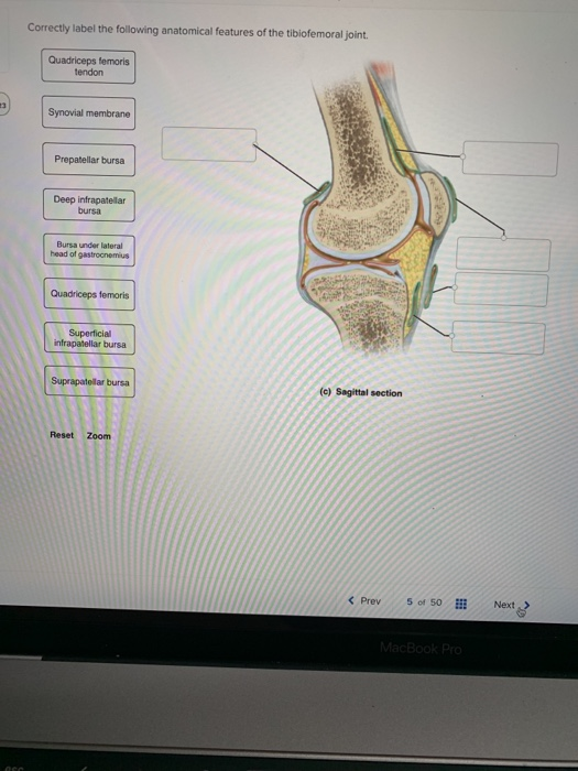
Solved Correctly label the following anatomical features ... Question: Correctly label the following anatomical features of the tibiofemoral joint. Articular cartilage Synovial membrane Joint capsule Quadriceps femoris Patellar ligament Femur Infrapatellar fat pad Tibia Quadriceps femoris tendon Meniscus Patella (c) Sagittal section Reset Zoom This problem has been solved! See the answer 9.1 Classification of Joints - Anatomy & Physiology An axis in anatomy is described as the movements in reference to the three anatomical planes: transverse, frontal, and sagittal. Thus, diarthroses are classified as uniaxial, biaxial, or multiaxial joints. A uniaxial joint only allows for a motion in a single plane (around a single axis). The elbow joint, which only allows for bending or ...
Correctly label the following anatomical features of the ... The tibiofemoral joint is the hinge synovial joint which joins the distal femur to proximal tibia. 2. Lateral condyle of femur- it is one of the projection on lateral side at lower extremity of femur. The lateral tibiofemoral joint is articulation between rounded lateral condyle of femur and relatively flat condyle of tibia. 3.
Correctly label the following anatomical features of the tibiofemoral joint.
module 6 quiz questions exam 2 Flashcards | Quizlet Correctly label the following anatomical features of the tibiofemoral joint. ATP and creatine phosphate are collectively known as the _____, which provides nearly al the energy required for short bursts of intense activity. Lab 7 Musculoskeletal Anatomy Part 3: Articulation and ... The system has the muscle pull is on the same side of the joint as the resistance placed on the joint but will have the resistance placed on the joint falls between the joint moving and muscle pull causing the movement, similar to the action of using a nutcracker. This type of lever is seen at the Tibiotalar and Talofibular (ankle) articulation. Knee Joint - Anatomy Pictures and Information The knee, also known as the tibiofemoral joint, is a synovial hinge joint formed between three bones: the femur, tibia, and patella. Two rounded, convex processes (known as condyles) on the distal end of the femur meet two rounded, concave condyles at the proximal end of the tibia. Continue Scrolling To Read More Below... Additional Resources
Correctly label the following anatomical features of the tibiofemoral joint.. Femur Bone Anatomy: Labeled Diagram, Quiz, Color-Coded ... There is a simple way to remember the main anatomical features of the femur using a human stick figure as drawn below. The different parts of the human stick figure correlate with different parts of the femur. Let's walk through the stick figure starting at the head (superior/proximal) and moving to the legs (inferior/distal). Label The Structures Of The Knee. : Label Diagram Of The ... Label the structures of the knee. The foot is an extremely complex anatomic structure made up of 26 bones and 33 joints that must work together with 19 muscles and 107 ligaments to execute . Start studying knee joint label. Anatomy of the knee including labeling . To deepen the articular surface of the tibia, . correctly label the following anatomical features of a ... Correctly label the following anatomical features of the tibiofemoral joint. Fibula Patellar ligament (cut) Patellar surface Femur Lateral condyle Medial condyle | Lateral meniscus Transverse ligament Tibia (a) Anterior view This is the bone of the upper leg. Drag the appropriate labels to their respective targets. AHCDWeek3SOL5.pdf - 5. Award: 10.00 points ... - Course Hero Correctly label the following parts of bone cells. Explanation: Osseous tissue consists of cells and matrix, like any other connective tissue, and the matrix consists of fibers and ground substance. There are four kinds of bone cells: osteogenic cells, osteoblasts, osteocytes, and osteoclasts.
PDF BIO 113 LAB 1. Anatomical Terminology, Positions, Planes ... Surface Anatomy . Body surfaces provide a number of visible landmarks that can be used to study the body. Several of these are described on the following pages. Locating Body Landmarks . Anterior Body Landmarks . Identify and use anatomical terms to correctly label the following regions on Figure 1: Proximal - TeachMeAnatomy - Making Anatomy Simple The proximal and distal tibiofibular joints refer to two articulations between the tibia and fibula of the leg. These joints have minimal function in terms of movement but play a greater role in stability and weight-bearing. In this article, we shall look at the anatomy of the proximal and distal tibiofibular joints - their structures, neurovascular supply and clinical relevance. Screenshot (175).png - 3 Correctly identify the following ... View Screenshot (175).png from SCIENCE 100-7 at Jersey College. 3 Correctly identify the following parts of a synovial joint. Fibrous capsule nts Joint capsule Proximal phalanx Joint cavity Chapter 9 QS Anatomy (Joints) Flashcards - Quizlet Correctly identify the following anatomical parts of the temporomandibular joint. external acoustic meatus, temporomandibular ligament, sphenomandibular ligament, styloid process, articular capsule The temporomandibular joint is limited to protraction and retraction due to its articular disc. False
Solved Correctly label the following anatomical features ... Question: Correctly label the following anatomical features of the tibiofemoral joint. Synovial membrane Femur Medial meniscus Patella Tibia Joint cavity Infrapatellar fat pad (e) Sagittal section Reset Zoom This problem has been solved! See the answer Show transcribed image text Expert Answer Anatomy of Selected Synovial Joints | Anatomy and Physiology I This section will examine the anatomy of selected synovial joints of the body. Anatomical names for most joints are derived from the names of the bones that articulate at that joint, although some joints, such as the elbow, hip, and knee joints are exceptions to this general naming scheme. Articulations of the Vertebral Column Knee joint: anatomy, ligaments and movements | Kenhub The tibiofemoral joint is an articulation between the tibia and the femur, while the patellofemoral joint is an articulation between the patella and the femur. The knee joint is the largest and arguably the most stressed joint in the body. Chapter 6 Anatomy Quiz Flashcards | Quizlet Joint _____ is maintained by the isometric contraction of antagonistic muscles at a single joint. ... Correctly label the following anatomical features of the tibiofemoral joint. Sets with similar terms. Chapter 9 muscle tissue. ... Features. Quizlet Live. Quizlet Checkpoint. Quizlet Learn. Explanations. Flashcards. Mobile.
Solved: Correctly Label The Following Anatomical Features ... | Chegg.com
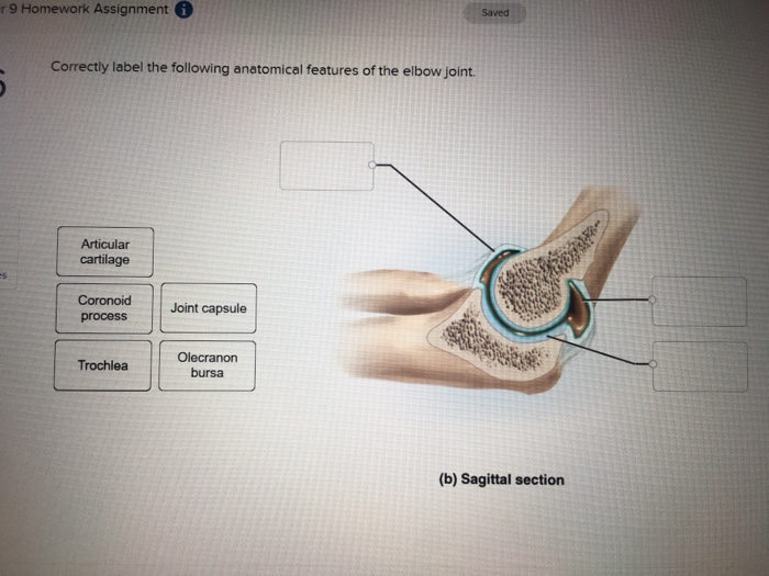
9.6 Anatomy of Selected Synovial Joints - Anatomy & Physiology Elbow Joint. The elbow joint is a uniaxial hinge joint formed by the humeroulnar joint, the articulation between the trochlea of the humerus and the trochlear notch of the ulna.Also associated with the elbow are the humeroradial joint and the proximal radioulnar joint. All three of these joints are enclosed within a single articular capsule (Figure 9.6.4).

33 Correctly Label The Following Anatomical Features Of The Tibiofemoral Joint. - Labels Design ...
Solved Correctly label the following anatomical features ... Correctly label the following anatomical features of the tibiofemoral joint. Fibula Patellar ligament (cut) Patellar surface Femur Lateral condyle Medial ...1 answer · Top answer: Description of parts- 1. Femur- It is the upper bone of the leg and largest bone of the body. The head forms the ball and socket joint with the ...
Solved: R 9 Homework Assignment Saved Correctly Label The ... | Chegg.com
Synovial Joints | Anatomy and Physiology I The joint with the greatest range of motion is the ball-and-socket joint. At these joints, the rounded head of one bone (the ball) fits into the concave articulation (the socket) of the adjacent bone (see Figure 3f). The hip joint and the glenohumeral (shoulder) joint are the only ball-and-socket joints of the body.

33 Correctly Label The Following Anatomical Features Of The Tibiofemoral Joint. - Labels Design ...
Module Five- Joints Flashcards - Quizlet Bones that join together and are held in place with threads of collagen form a joint that is called a (n) synarthrosis. 3. Bones joined together with cartilage between the ends of the bones form a joint called a (n) amphiarthrosis. 4. The most complex joints are called synovial joints. They display varying amounts of mobility. Suture joint

34 Correctly Label The Following Anatomical Features Of The Tibiofemoral Joint. - Labels Design ...
Bio quiz 6 Flashcards | Quizlet With low-frequency stimulation, the muscle relaxes fully between contractions, resulting in identical twitches per stimulus. 2. As the frequency gets progressively higher, the muscle does not have time to relax in between twitches. Each twitch rides "piggyback" on the previous one and generates higher tension. 3.

Solved: Correctly Label The Following Anatomical Features ... | Chegg.com
The Knee Joint - TeachMeAnatomy - Making Anatomy Simple The knee joint is a hinge type synovial joint, which mainly allows for flexion and extension (and a small degree of medial and lateral rotation). It is formed by articulations between the patella, femur and tibia. In this article, we shall examine the anatomy of the knee joint - its articulating surfaces, ligaments and neurovascular supply.

34 Philip Phillips Record Label - Online Labels Ideas
Solved Correctly label the following anatomical features ... Question: Correctly label the following anatomical features of the tibiofemoral joint. Tibial collateral li Fibular Anterior cruciate ligament Femur Tibia (cut) (a) Anterior view This problem has been solved! See the answer Show transcribed image text Expert Answer 96% (23 ratings)
Solved: Correctly Label The Following Anatomical Features ... | Chegg.com
Free Science Flashcards about ANP1040 Exam 3 - StudyStack Correctly label the following anatomical features of the tibiofemoral joint. Tibia, Femur, Medial Condyle, Lateral Condyle, Anterior Cruciate Ligament, Posterior Cruciate Ligament, Patellar Surface, Fibula: Correctly label the following anatomical features of connective tissue of muscle.
0 Response to "44 correctly label the following anatomical features of the tibiofemoral joint."
Post a Comment