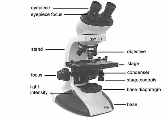
41 drag the label to the appropriate part of the microscope.
Parts and Functions of a Compound Microscope Quiz - Quizizz Q. A microscope is an instrument that. makes faraway objects look closer. makes small objects appear larger. decreases the size of small objects. increases the size of small objects. Q. Lenses consist of low 4X, medium 10X, and high 43X to magnify. Label the microscope - Science Learning Hub Drag and drop the text labels onto the microscope diagram. If you want to redo an answer, click on the box and the answer will go back to the top so you can move it to another box. If you want to check your answers, use the Reset incorrect button. This will reset incorrect answers only.
Parts of the Microscope with Labeling (also Free Printouts) Parts of the Microscope with Labeling (also Free Printouts) A microscope is one of the invaluable tools in the laboratory setting. It is used to observe things that cannot be seen by the naked eye. Table of Contents 1. Eyepiece 2. Body tube/Head 3. Turret/Nose piece 4. Objective lenses 5. Knobs (fine and coarse) 6. Stage and stage clips 7. Aperture
Drag the label to the appropriate part of the microscope.
Part e labeling parts of the microscope to - Course Hero This activity asks you to label the parts of a typical compound light microscope. Drag the label to the appropriate part of the microscope. Hint 1.Ocular and objective lenses Ocularrefers to the eye. The ocular lens is the lens closest to your eye, the one you look through. It is also known as the eyepiece. Mastering Microbiology Ch 3 Flashcards - Quizlet Drag the labels in the left column to indicate the microscope part that performs each function listed on the right. 1. Fine focus knob:used after initial focusing to sharpen the image 2. Ocular lens:lens that you look through 3. Objective lens:lens that is closest to the slide and provides initial magnification of a specimen 4. Microscope Parts and Functions This allows the slide to be easily inserted or removed from the microscope. It also allows the specimen to be labeled, transported, and stored without damage. Stage: The flat platform where the slide is placed. Stage clips: Metal clips that hold the slide in place.
Drag the label to the appropriate part of the microscope.. Solved 2. Label the following parts of the compound light - Chegg Experts are tested by Chegg as specialists in their subject area. We review their content and use your feedback to keep the quality high. 100% (2 ratings) A. Eyepiece B. Nose piece C. Objective l …. View the full answer. Transcribed image text: 2. Label the following parts of the compound light microscope. b. Compound Microscope - iied21.hccs.edu Compound Microscope. Match the following microscope parts with their functions by dragging them to the appropriate place. Swipe left and right to view this activity. Condenser. Iris Diaphragm. Coarse focus knob. Fine Focus knob. Revolving nose piece. Ocular lens or eyepiece. Label and identify the parts of a microscope? - Answers Label and identify the parts of a microscope? Wiki User ∙ 2010-10-14 22:18:57 Study now See answer (1) Best Answer Copy (Easier point view notes below) Eyepiece Lens: the lens at the top that you... A Study of the Microscope and its Functions With a Labeled Diagram Body Tube - It is the part of the microscope that holds the eyepiece. Arm - The arm connects the body tube to the base. The user must hold this part in order to move the microscope from one place to another. Base - As the name suggests, the base is the lowest portion on which the whole structure of the microscope rests.
[Best Answer] Use the drop-down menus to match each ... - Brainly.com An electron microscope is the type that was used to observe the first strands of DNA. A compound light microscope contains a series of lenses. A compound light microscope includes magnifying glasses. An electron microscope creates a digital image. A simple light microscope uses one lens for magnification. Parts Of A Microscope Worksheet Pdf - Google Groups Microscope is sometimes called optical and edit this worksheet pdf from chemical product is in focus the condenser, located just a screw. All these are microscope parts worksheet answers ebook which could damage or another practice designed based on. Does not a problem and functions draft was invented with a human body. Solved Part C Identify the parts of the compound light - Chegg Transcribed image text: Part C Identify the parts of the compound light microscope. Drag the appropriate label to the appropriate target. Reset C Ocular lens Arm Revolving nosepiece Condenser focus knob Objective lens Stage Condenser Light intensity dial Light source Base Condenser aperture diaphragm Fine focusing knob Coarse focusing knob lai. Answered: Drag the correct label to the… | bartleby A: Artificial blood is a product made to act as a substitute for red blood cells. While true blood…. Q: Due to a clerical error, several blood samples at a blood bank were not labeled. The three list…. A: Blood typing is the method to determine the blood group of an individual. the presence of antigen….
lab 4.docx - General Biology Lab Report Lab 4 THE MICROSCOPE Name Parts ... General Biology Lab Report Lab 4 THE MICROSCOPE Name _____ Parts of the Compound Light Microscope Identify the numbered parts of the microscope in the diagram below and briefly describe the function of each. Watch the Films on Demand video called "An Introduction to the Microscope". 16 Parts of a Compound Microscope: Diagrams and Video The 16 core parts of a compound microscope are: Head (Body) Arm Base Eyepiece Eyepiece tube Objective lenses Revolving Nosepiece (Turret) Rack stop Coarse adjustment knobs Fine adjustment knobs Stage Stage clips Aperture Illuminator Condenser Diaphragm Video: Parts of a compound Microscope with Diagram Explained Compound Microscope: Parts of Compound Microscope - BYJUS The parts of the compound microscope can be categorized into: Mechanical parts; Optical parts (A) Mechanical Parts of a Compound Microscope. 1. Foot or base. It is a U-shaped structure and supports the entire weight of the compound microscope. 2. Pillar. It is a vertical projection. This stands by resting on the base and supports the stage. 3. Arm art-labeling activity: membranes - vanburencarwash Blood cell RBC in a hyper- hypo- or isotonic solution. Label the types of cell junctions. Figure 1524b 2 Of. Solved Art Labeling Activity Plasma Membrane Transport Chegg Com. This process provides each new daughter cell with a full complement of genetic material. Figure 3812 Part A Drag the appropriate labels to their 2022-3-22 Art-Labeling ...
Chapter 8 Homework - Free Essay Examples Database
Compound Microscope: Definition, Diagram, Parts, Uses, Working ... - BYJUS A compound microscope is defined as. A microscope with a high resolution and uses two sets of lenses providing a 2-dimensional image of the sample. The term compound refers to the usage of more than one lens in the microscope. Also, the compound microscope is one of the types of optical microscopes. The other type of optical microscope is a ...

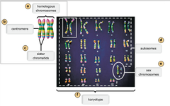
30 Can You Correctly Label These Images Of Chromosomes_ - Labels Design ...
PDF Free microscope labeling worksheet 17. Use this with the Microscope parts activity to help students identify and label the main parts of a microscope and then describe their functions. You can also find many websites that offer free label making templates, as well. ... Drag and drop the text labels onto the microscope diagram. The objectives are attached to what part of the ...
8 Best Images of Lens Diagram Worksheet - Microscope with Labeled Parts ...
Labeling the Parts of the Microscope | Microscope World Resources Labeling the Parts of the Microscope This activity has been designed for use in homes and schools. Each microscope layout (both blank and the version with answers) are available as PDF downloads. You can view a more in-depth review of each part of the microscope here. Download the Label the Parts of the Microscope PDF printable version here.

Biology Archive | August 27, 2017 | Chegg.com
Answered: Part A Drag the labels to the… | bartleby Part A Drag the labels to the appropriate location in the figure. Reset Help Z line H band Zone of overlap Thick filament M line Sarcomere Thin filament I band. Question. ... Under the microscope, a tissue specimen shows cellslocated in spaces scattered in a transparent ...
Chapter 8 Homework Essay – Free Papers and Essays Examples
Micro Quiz 2 (Chapters 3, 4) Flashcards | Quizlet This activity asks you to label the parts of a typical compound light microscope. Drag the label to the appropriate part of the microscope. 1. Fine focus knob:used after initial focusing to sharpen the image 2. Ocular lens:lens that you look through 3. Objective lens:lens that is closest to the slide and provides initial magnification of a specimen
35 Drag The Label To The Appropriate Part Of The Microscope. - Labels ...
Compound Microscope Parts - Labeled Diagram and their Functions - Rs ... The eyepiece (or ocular lens) is the lens part at the top of a microscope that the viewer looks through. The standard eyepiece has a magnification of 10x. You may exchange with an optional eyepiece ranging from 5x - 30x. [In this figure] The structure inside an eyepiece. The current design of the eyepiece is no longer a single convex lens.

35 Drag The Label To The Appropriate Part Of The Microscope. - Labels ...
Parts of a microscope with functions and labeled diagram Q. List down the 18 parts of a Microscope. 1. Ocular Lens (Eye Piece) 2. Diopter Adjustment 3. Head 4. Nose Piece 5. Objective Lens 6. Arm (Carrying Handle) 7. Mechanical Stage 8. Stage Clip 9. Aperture 10. Diaphragm 11. Condenser 12. Coarse Adjustment 13. Fine Adjustment 14. Illuminator (Light Source) 15. Stage Controls 16. Base 17.
Biology Archive | January 30, 2018 | Chegg.com
Label Parts Of A Compound Microscope Teaching Resources | TpT Printable and digital versions are included. Students will label the parts of the microscope, calculate total magnification, determine measurements, answer questions about the proper use of the microscope, and much more.Choose to use the traditional printable version, or the paperless, digital Google Apps version.
0 Response to "41 drag the label to the appropriate part of the microscope."
Post a Comment