43 how to label gel electrophoresis images
CHAPTER 10 Flashcards | Quizlet Please label the images to review the process of polymerase chain reaction and how its products can be analyzed using gel electrophoresis. Match the components of a typical PCR reaction with the function they serve. ... Please label the images to review the process of screening bacterial clones for those containing a donor gene. Other sets by ... How to Prepare an Electrophoresis Argarose Gel - Instructables Make sure to label each stock solution appropriately. If you already have these prepared in your lab go to the next step. 1) Prepare 1X TAE Stock Solution a. Pour 20 ml of 50X TAE solution into 1 liter plastic bottle b. Bring final volume to 1 liter with distilled water c. Gently shake solution NOTE : Many labs have 50X TAE solution on hand.
Solved Please label the images to review the process of - Chegg Expert Answer Answer) The complete DNA molecules with primer and buffer containing nucleotides and taq polymer … View the full answer Transcribed image text: Please label the images to review the process of polymerase chain reaction and how its products can be analyzed using gel electrophoresis.
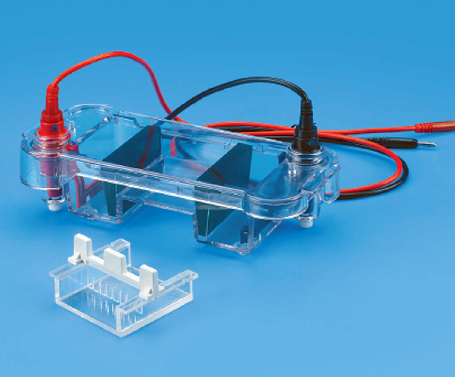
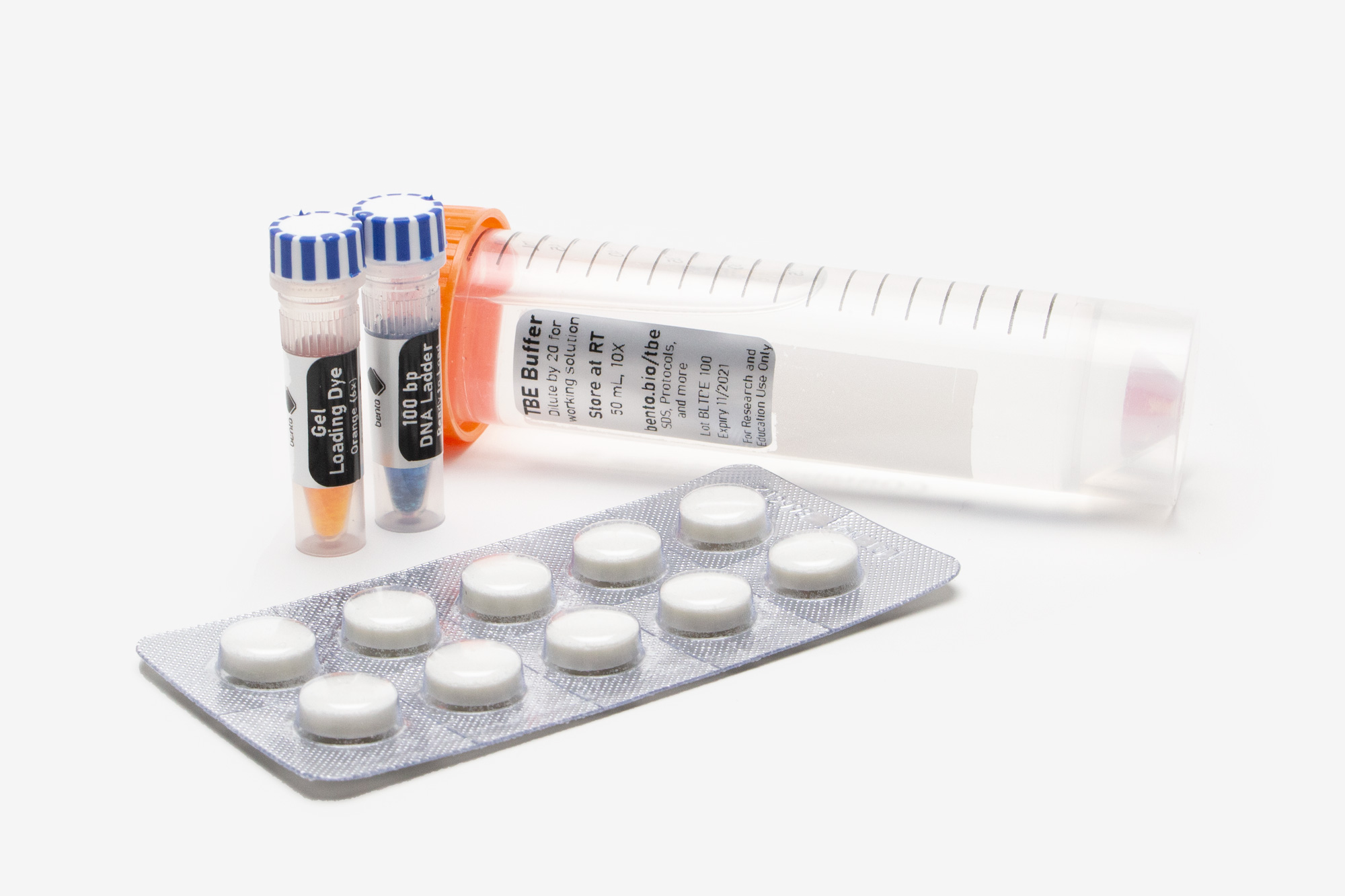
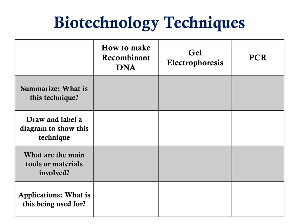
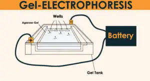
How to label gel electrophoresis images
Solved Please label the images to review the process of - Chegg Question: Please label the images to review the process of polymerase chain reaction and how its products can be analyzed using gel electrophoresis. Dam Deration Denaturation 1 се DNA Replication Pricing Olgorde sha and of of arcon A Cole 770 Restriction andonucleases selectively cleaving sites of DNA cony Piring w Opelweg () Restriction ... ChemiDoc Imaging Systems | Bio-Rad ChemiDoc Imagers offer best-in-class performance with ease of use for visible light (RGB) and far red/near infrared (FR/NIR) fluorescence and chemiluminescence detection and all general gel documentation applications.Stain-free imaging enables immediate visualization of proteins without gel staining and instant verification of protein transfer to blots. How to quantify each band in gel electrophoresis? - ResearchGate you can do an analytical curve in a 1d gel, with known amounts of bsa for example, use photoshop to quantify the pixels (the curve would be pixels x protein mass you applied for each well) and then...
How to label gel electrophoresis images. THEV1 - Overview: Thalassemia and Hemoglobinopathy … This evaluation will always include hemoglobins A2 and F and hemoglobin electrophoresis utilizing cation exchange high-performance liquid chromatography (HPLC) and capillary electrophoresis methods. If a serum sample is received, a serum ferritin will always be performed to allow incorporation of possible iron deficiency into profile interpretation and … Polyacrylamide Gel Electrophoresis - Cleaver Scientific The gel mixture is made up not in water but in electrophoresis buffer (Tris-HCl), that provides the ions for electrophoresis. Often, the gel is poured in 2 parts. The first parts is a resolving gel, with a pH around 8.8 which slows the migration of the proteins. Above the resolving gel, a stacking gel is poured with a pH of 6.8 and a larger ... How to Interpret DNA Gel Electrophoresis Results - GoldBio During gel electrophoresis, you may have to load uncut plasmid DNA, digested DNA fragment, PCR product, and probably genomic DNA that you use as a PCR template into the wells. Your digested DNA fragment is a digested PCR product. The next step is to identify those bands to figure out which one to cut. Gel Electrophoresis. Lane 1: DNA Ladder. Stain-Free Imaging Technology | Bio-Rad Stain-Free imaging technology utilizes a polyacrylamide gel containing a proprietary trihalo compound to make proteins fluorescent directly in the gel with a short photoactivation, allowing the immediate visualization of proteins at any point during electrophoresis and western blotting. This trihalo compound is covalently bound to tryptophan residues, enhancing their …
How to make a gel image using Powerpoint - YouTube A quick tutorial on how to make a reasonably polished figure using an image of a gel using Powerpoint. There are certainly more professional ways of doing th... Annotating A Gel | Get Your Science On Wiki | Fandom Part 1. Photo Editing: 1.Take your JPG or PNG file of your Gel and open it with a photo editing program (GIMP). 2. Under "Image" --> "Transform" rotate your picture by 90 degrees so that your wells are on top of the page. 3. Using the Crop tool Cut out the black borders leaving only the gel. 4. gel electrophoresis | Learn Science at Scitable - Nature In gel electrophoresis, the molecules to be separated are pushed by an electrical field through a gel that contains small pores. The molecules travel through the pores in the gel at a speed that ... Methods for Labeling Nucleic Acids - Thermo Fisher Scientific Typically, nucleic acids hybridization reactions (i.e., northern blotting) benefit from the high specific activity gained through random incorporation of label into a probe. However, assays requiring protein interactions (i.e., gel shift and pull-down assays) require end-labeling to allow protein binding.
EPU - Overview: Electrophoresis, Protein, 24 Hour, Urine PEU: Agarose Gel Electrophoresis. IFXU: Immunofixation. NY State Available. Indicates the status of NY State approval and if the test is orderable for NY State clients. Yes Reporting Name. Lists a shorter or abbreviated version of the Published Name for a test Electrophoresis, Protein, 24 Hr, U Aliases. Lists additional common names for a test, as an aid in searching Bence … PDF Gel Electrophoresis Size Marker - dia-m.ru Labeling in four positions via the terminal EcoR I generated recessed ends is possible, especially with the DNA ladder 100 bp (A3470) and the DNA Ladder Mix 100 - 5000 (A3660). For the labeling of the DNA, the product is simply dissolved in TE buffer or bidistilled water. DNA staining with methylene blue Analysis of protein gels (SDS-PAGE) - Rice University Calibrate the gel using standards of known molecular mass (set up a standard curve if necessary) Select polypeptide bands in the lane (s) of interest to be analyzed and identify them by some generic label (e.g., a, b, c,... or 1, 2, 3,...) Estimate molecular mass or relative molecular mass for each band of interest Agarose Gel Electrophoresis for the Separation of DNA Fragments Place an appropriate comb into the gel mold to create the wells. Pour the molten agarose into the gel mold. Allow the agarose to set at room temperature. Remove the comb and place the gel in the gel box. Alternatively, the gel can also be wrapped in plastic wrap and stored at 4 °C until use ( Fig. 1 ). 2.
Gel electrophoresis (article) - Khan Academy When a gel is stained with a DNA-binding dye and placed under UV light, the DNA fragments will glow, allowing us to see the DNA present at different locations along the length of the gel. The bp next to each number in the ladder indicates how many base pairs long the DNA fragment is. A well-defined "line" of DNA on a gel is called a band.
Typhoon™ laser-scanner platform | Cytiva Typhoon™ is the platform of choice to support efficiency by reducing the risk of unexpected HCP.Typhoon™ mitigates HCP risk through 2D DIBE coverage analysis and approach bridging studies that provide more confidence in late clinical trial phases.. The right filter and laser configuration minimize cross talk between wavelength channels.
Gel Electrophoresis: Basics & Steps - SchoolWorkHelper Basic Steps. Aragonese and the buffer are mixed together and microwaved to create the gel. It is poured into a mold and has a "comb" placed in it to make holes for the DNA to be inserted. Once it has cooled the comb is removed. The gel is then placed in the gel electrophoresis box and buffer solution is poured onto it.
Analysis of protein gels (SDS-PAGE) - Rice University Calibrate the gel . The stacking gel is of no use to the analysis and it can be removed. Top of the gel refers to the top of the separating gel, that is, the point at which different polypeptides began to separate. A mix of protein standards usually consists of five to eight individual polypeptides that produce a prominent "ladder." Standards ...
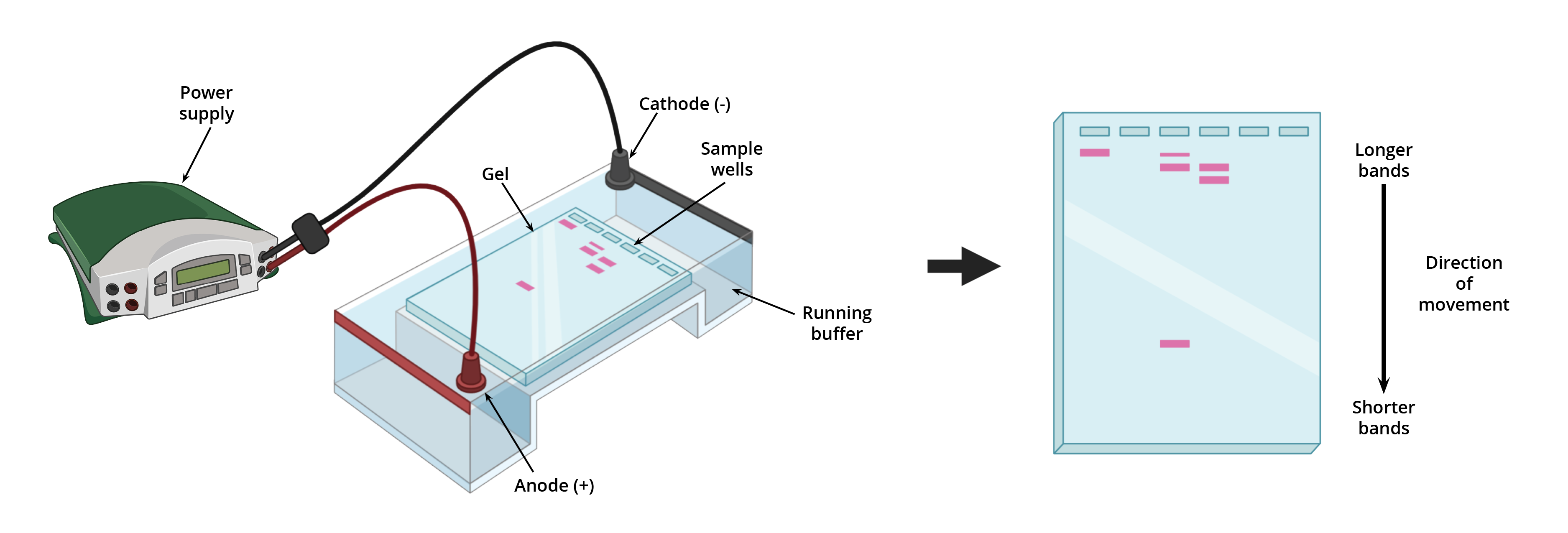
Gel Electrophoresis: Definition, Principle, and Application Electrophoresis is a process used for the separation of macro and micro molecules in an electric field by applying charges at both the extents. The mixture of substances is spread in the supporting film. The supporting films are placed in a salt solution filled in a container, where one container holds a cathode and the other carries an anode.
Analyzing gels and western blots with ImageJ - lukemiller.org After drawing the rectangle over your first lane, press the 1 (Command + 1 on Mac) key (Command + 1 on Mac) or go to Analyze>Gels>Select First Lane to set the rectangle in place. The 1st lane will now be highlighted and have a 1 in the middle of it. 5.
3 Ways to Read Gel Electrophoresis Bands - wikiHow Hold a UV light up to the gel sheet to reveal results when using a UV-based dye. With your gel sheet in front of you, find the switch on a tube of UV light to turn it on. Hold the UV light 8-16 inches (20-41 cm) away from the gel sheet. Illuminate the DNA samples with the UV light to activate the dye and read the results.
PDF Lab 4: Gel Electrophoresis - Vanderbilt University This protocol uses a standard electrophoresis system. The agarose gel will be made by adding agarose powder (or tablets) to running buffer, boiling the mixture, then letting it cool into a gelatin-like slab. The agarose gel is run in a standard electrophoresis system, then visualized with a transilluminator. Pre-Lab Preparation
Gel Electrophoresis - an overview | ScienceDirect Topics For gel electrophoresis, a DNA sample is loaded at one end of a gel matrix (usually agarose or acrylamide) that provides a uniform pore size through which the DNA molecules can move. Application of a constant electric field causes DNA fragments (all have a uniform, strong negative charge) to migrate toward the cathode.
Smearing in agarose gel of PCR product? - ResearchGate Primer dimers (PDs) formed during PCR run is a common finding which be visible after gel electrophoresis of the PCR product. PDs in ethidium bromide …
GelAnalyzer GelAnalyzer 19.1 Analyze gel images from any source Use your digital camera, smartphone, or gel doc system to obtain images. GelAnalyzer will take care of the rest. Automatic lane and band detection With full manual control over adding, modifying, and deleting lanes and bands. Fix run distortions through Rf calibration
Lysyl Endopeptidase®, Mass Spectrometry Grade (Lys-C) Among the most important techniques in proteome analyses is the in-gel digestion of protein spots/bands that have been resolved by electrophoresis using digestive enzymes, such as trypsin and lysyl endopoptidase. Proteins can be identified by mass spectrometry analysis of the peptides produced by in-gel digestion, and further information regarding post-translational …
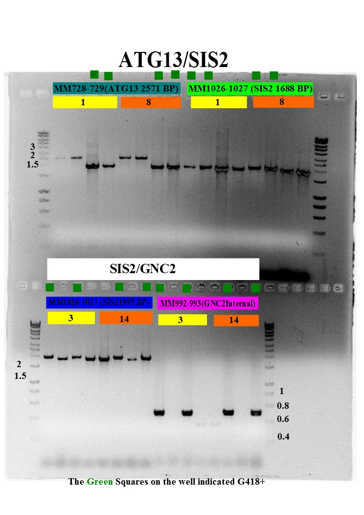
A Complete Guide for Analysing and Interpreting Gel Electrophoresis Results let see some of the gel images of PCR fragments. 2% gel is required to separate PCR products because PCR products are the smaller fragments of DNA nearly ~100bp to ~1500bp. Image 1: The image is captured under the UV transilluminator instead of the gel doc system to show you the effect of EtBr on the gel electrophoresis results.
PDF Gel Electrophoresis: How Does It Work - Purdue University a. After you find out what dyes you are using, draw a picture of the gel and the wells. Label which dyes you will put in each well. b. When you load a gel, it is very important that you do not damage the gel in any way. You must be very careful not to "jab" the gel with the end of your pipet. Ideally, you shouldn't even touch the gel with the ...
ImageJ for Editing & Labelling PCR Gel Image - YouTube This Tutorial is all about how to quickly Edit & Label PCR Gel Image Using ImageJ software. Presented by - Elvis SamuelJoin Our Telegram Channel for free Sof...
Part 2: Analyzing and Interpreting (Agarose) Gel Electrophoresis Results Now let see some real images. The gel image above is the result of restriction digestion. Lane 3, 5, 7, and 8 are a homozygous normal allele with a 184bp band here one band of 68bp is also present, but it is not visible. Lane 2 is a mutant uncut allele of 252bp. Lane 1 and 6 are heterozygous contain three alleles: 252bp, 184bp and 68bp.
How can I modify a photograph of gel electrophoresis taken with ... After amplifying my target gene (1900bp) into the required cDNA by PCR,>cut specific gel bands and purify the gel and the concentration was 30ng/ul. Next, performed A-Tailing according to the Kit...
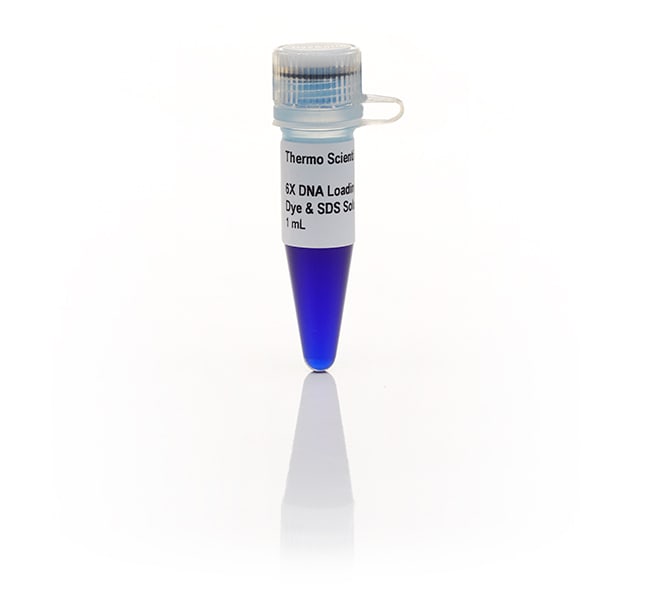
Agarose gel electrophoresis of DNA - Cleaver Scientific For a typical agarose gel electrophoresis procedure, the gel matrix is cast as a horizontal slab. Plastic combs are used to create indentations, or wells, into which the DNA is loaded. Before loading, the DNA is mixed with a loading dye that weighs down the sample in the solution, so it does not leave the well, and also includes a visible marker to track the progression of the run.





0 Response to "43 how to label gel electrophoresis images"
Post a Comment