40 label the spinal cord meninges and spaces.
en.wikipedia.org › wiki › Arachnoid_granulationArachnoid granulation - Wikipedia Arachnoid granulations (also arachnoid villi, and pacchionian granulations or bodies) are small protrusions of the arachnoid mater (the thin second layer covering the brain) into the outer membrane of the dura mater (the thick outer layer). (Get Answer) - Art-labeling Activity: The spinal meninges and ... Art-labeling Activity: The spinal meninges and associated structures Adipose tissue space Epidural.. Art-labeling Activity: The spinal meninges and associated structures Adipose tissue space Epidural space Dura mater Dorsal root ganglion Spinal cord Dorsal root Arachnoid mater Ventral root Pia mater. label the structure.
Drag The Labels Onto The Diagram Of The Cns Meninges Drag The Labels Onto The Diagram Of The Cns Meninges. Terms in this set (15) drag the labels onto the diagram to identify the gross anatomical structures of the spinal cord. Drag the labels onto the diagram to identify the stages of the cell. Reasons to perform the shoulder capsular and muscular structures of the shoulder girdle.

Label the spinal cord meninges and spaces.
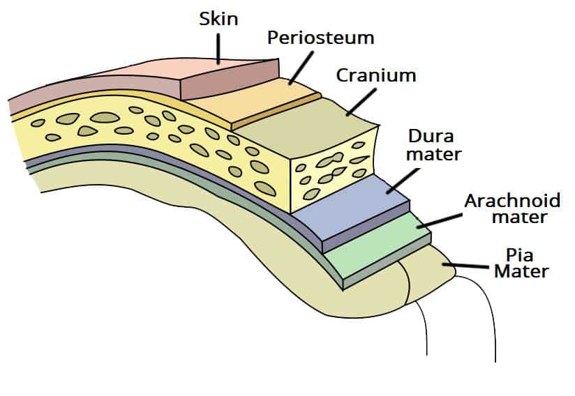
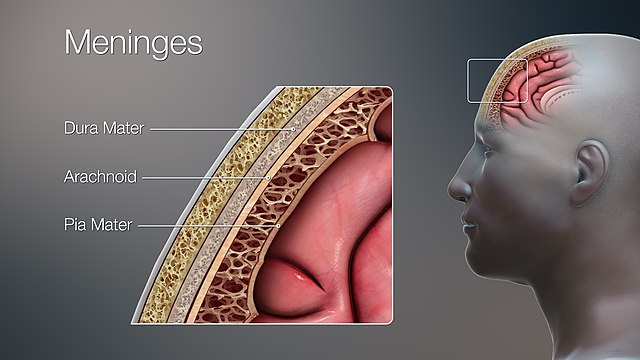
› image › nervovNervous System: Explore the Nerves with Interactive Anatomy ... Nov 02, 2020 · Each spinal nerve exits from the spinal cord through the intervertebral foramen between a pair of vertebrae or between the C1 vertebra and the occipital bone of the skull. Meninges The meninges are the protective coverings of the central nervous system (CNS). The Meninges - Dura - Arachnoid - Pia - TeachMeAnatomy The meninges refer to the membranous coverings of the brain and spinal cord. There are three layers of meninges, known as the dura mater, arachnoid mater and pia mater. These coverings have two major functions: Provide a supportive framework for the cerebral and cranial vasculature. Meninges Layers, Function & Anatomy - Study.com The brain and spinal cord are important structures that control the nervous system. These organs are covered by layers of tissues called meninges. There are three layers of meninges: Dura Mater...
Label the spinal cord meninges and spaces.. sciencetrends.com › anatomical-body-planesAnatomical Body Planes | Science Trends Jan 01, 2019 · The dorsal cavity is one long continuous cavity that houses portions of the central nervous system including the spinal cord and brain. It is found on the body’s dorsal side. The cranial cavity contains the cerebrospinal fluid, the brain’s meninges, and the brain itself. The cranial cavity overlaps with the anterior portion of the dorsal ... Neuroanatomy, Cranial Meninges - StatPearls - NCBI Bookshelf The brain and spinal cord are enveloped within three layers of membrane collectively known as the meninges, with the cranial meninges specifically referring to the section that covers the brain. From superficial to deep, the three layers are the dura, arachnoid, and pia—the term "mater," Latin for mother, often follows these names (i.e., dura mater, arachnoid mater, pia mater).[1] The ... Chapter 14 Assignment Flashcards | Quizlet WebInterventricular foramen 10. Lateral ventricle, The meninges is a three-layered, membranous covering of the brain and spinal cord. Read the descriptions below and then click and drag them into the appropriate box based on whether they pertain to the dura mater, the arachnoid mater, or the pia mater. 1. Dura Mater 2. Arachnoid Mater 3. Pia … Brain, Spinal Cord, Meninges Flashcards | Quizlet Purple: Spinal Pia Mater Label the spinal meninges and spaces enlarged subarachnoid space, ends at S2 vertebral level Lumbar Cistern - Lateral extensions of pia materu000b - Pass through arachnoid and attach internal surface to dura materu000b - Function to anchor the spinal cord Denticulate Ligaments Denticulate ligaments
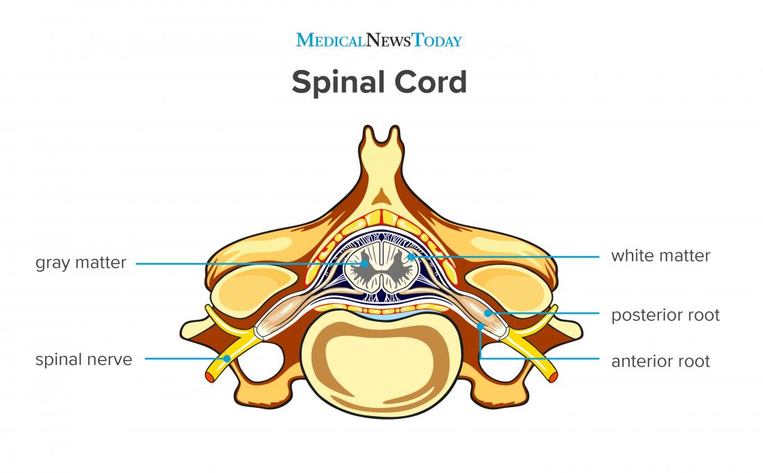
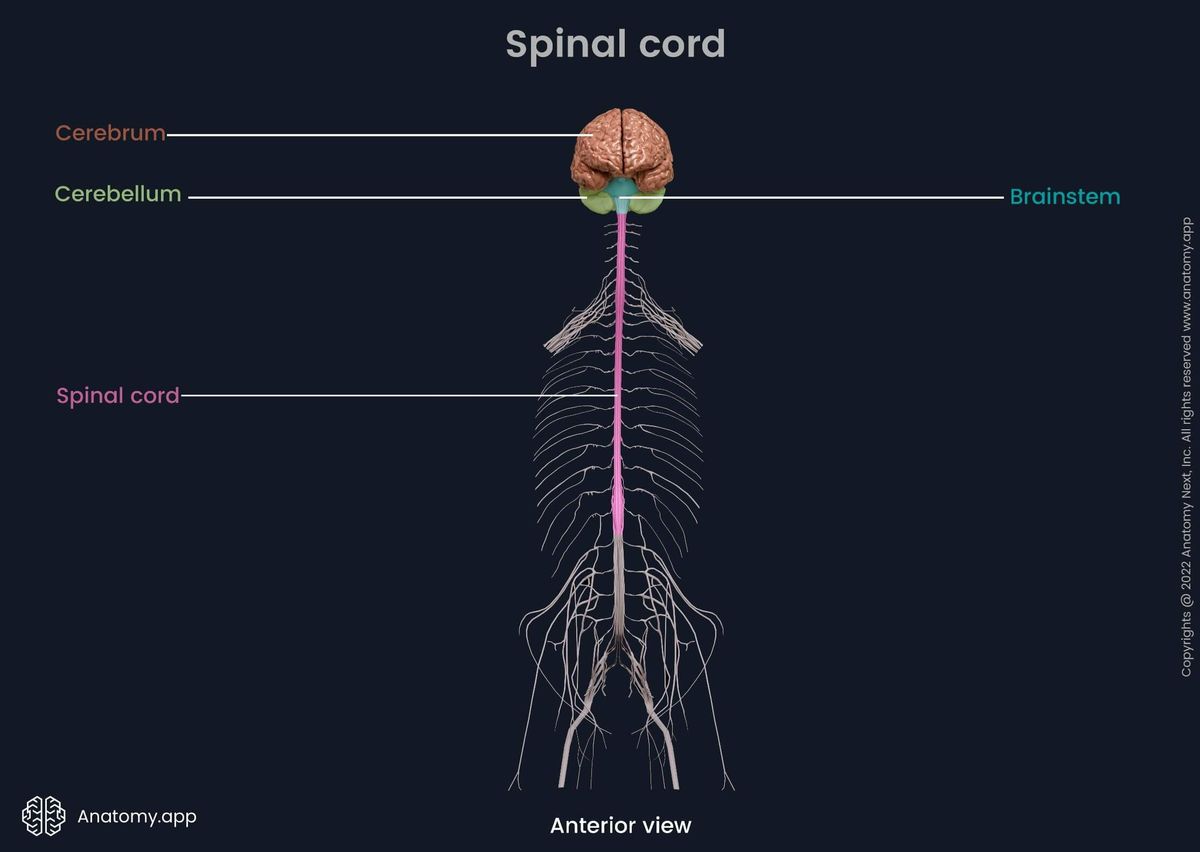
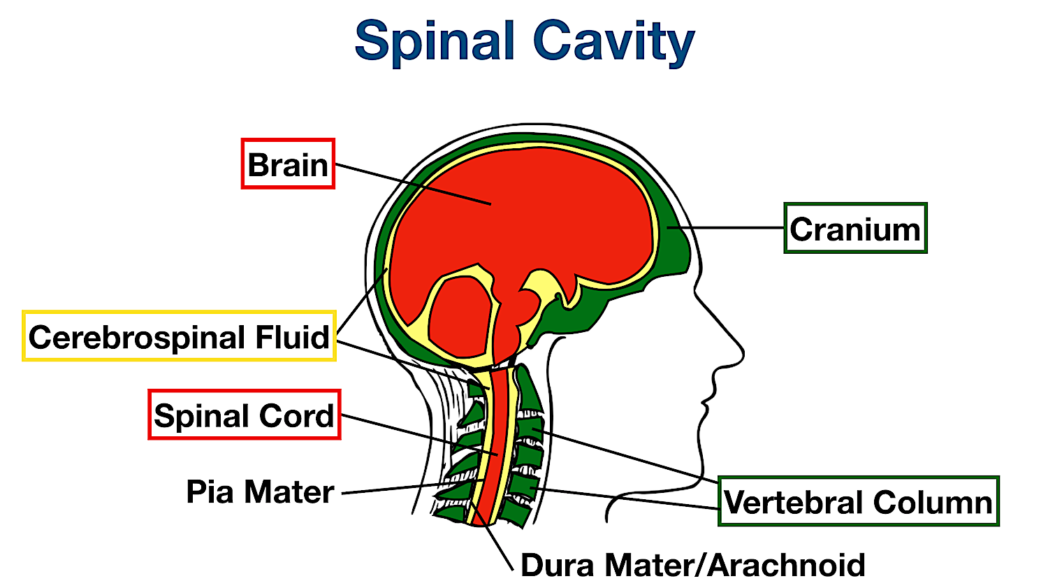
Spinal cord - Wikipedia The spinal cord is a long, thin, tubular structure made up of nervous tissue, which extends from the medulla oblongata in the brainstem to the lumbar region of the vertebral column (backbone). The backbone encloses the central canal of the spinal cord, which contains cerebrospinal fluid.The brain and spinal cord together make up the central nervous system (CNS). Spinal Cord - Anatomy, Structure, Function, & Diagram - BYJUS Three layers of meninges surround the spinal cord and spinal nerve roots. Dura mater Arachnoid mater Pia mater Dura mater consists of two layers- periosteal and meningeal. Epidural space is present between the two layers. Subarachnoid space lies between the arachnoid mater and pia mater. It is filled with cerebrospinal fluid. Spinal Cord Injuries DiFiore's Atlas of Histology with Functional Correlations (11th … WebThere is shortage of references in higher teaching institutions especially in newly opened institutions engaged in training of various Veterinary professionals in the country. Brain Tumors: Types, Causes, 13 Warning Signs, Treatment Web27/04/2022 · Spinal tap: Your doctor may remove a sample of cerebrospinal fluid (the fluid that fills the spaces in and around the brain and spinal cord). This procedure is performed with local anesthesia. The doctor uses a long, thin needle to remove fluid from the lower part of the spinal column. A spinal tap takes about 30 minutes. You must lie flat for several …
Meninges: Dura, arachnoid, pia, meningeal spaces | Kenhub The meningeal spaces are the spaces between the meningeal layers. There are three clinically significant meningeal spaces; epidural, subdural, and subarachnoid. We have described the anatomy of each of the spaces in the text above, however, we'd like to recap the most important facts and sum them up in the following paragraphs. Epidural space Spinal cord meninges labeling Diagram | Quizlet Spinal cord meninges labeling STUDY Terms in this set (...) Arachnoid mater Dura mater subdural space Subarachnoid space Denticulate ligament Pia mater YOU MIGHT ALSO LIKE... Meninges & Blood Vessels of the Brain - Neuroanatomy | Kenhub Anatomy Guide Kenhub $14.99 STUDY GUIDE The Brain, Spinal Cord, and Meninges | Basicmedical Key At autopsy, the brain of a 35-year-old man with history of AIDS shows diffuse edema and opacification of the leptomeninges. On histologic examination, the subarachnoid space is filled with the organisms seen in this GMS-stained section and a scanty mononuclear infiltrate. The organism has a thick PAS-positive capsule. Nervous System: Explore the Nerves with Interactive Anatomy … Web02/11/2020 · Newly created CSF flows through the inside of the brain in hollow spaces called ventricles and through a small cavity in the middle of the spinal cord called the central canal. CSF also flows through the subarachnoid space around the outside of the brain and spinal cord. CSF is constantly produced at the choroid plexuses and is …
Use the diagram to label: - ppt download
J code list and How to Bill J Codes Correctly by the “UNITS” with ... WebC79.32 Secondary malignant neoplasm, cerebral meninges C81.00-C81.49 Hodgkin lymphoma C82.00-C82.89 Follicular lymphoma and other types of follicular lymphoma C82.90-C82.99 Follicular lymphoma, unspecified C83.00-C83.89 Non-follicular lymphoma C84.01-C84.09 Mycosis fungoides C84.10-C84.19 Sezary’s disease C84.40- C84.49 …

Anatomy of the vertebral canal | Osmosis
Lab 1: Vertebral column, spinal cord, spinal nerves, and back muscles ... 3 Identify the spinal cord, spinal nerves, and spinal meninges, and describe their relationships to the vertebral column. 4 Trace motor and sensory pathways to and from the spinal cord through spinal nerves. 5 Identify the erector spinae and splenius muscles of the back. VERTEBRAL COLUMN Vertebral Column as a Whole 1

Brain, Spinal Cord, Meninges Flashcards | Quizlet
(PDF) LIBRO PARA COLOREAR NETTER - Academia.edu WebBackground: The aim of our study was to examine the effect of mild maternal hypothyroidism on the apoptosis of the oocytes in the ovaries of rats in the early postnatal period during formation of oocytes and follicles.

Anatomy of the spinal cord - e-Anatomy
Spinal cord: Anatomy, structure, tracts and function | Kenhub The spinal cord and spinal nerve roots are wrapped within three layers called meninges. The outermost is the dura mater, underneath it is the arachnoid mater, and the deepest is the pia mater. Dura mater has two layers (periosteal and meningeal), between which is the epidural space.
The Meninges - Dura - Arachnoid - Pia - TeachMeAnatomy
Ventricles and Meninges | SchoolWorkHelper Science. Ventricles and Meninges. Ventricles= expansions of the brain's central cavity. Filled with CSF, lined with ependymal cells. Continuous with each other, central canal, and spinal cord. 1 st /2 nd = paired lateral ventricles (lie in cerebral hemispheres) [separated by septum pellucidum - transparent wall]

Spinal cord meninges labeling Diagram | Quizlet
Untitled Document [faculty.collin.edu] Lecture Review Spinal Cord & Spinal Nerves Dr. Weis 1. and specific protection provided. 2. Name all the meninges and spaces and functions 3. spinal nerve (#) 4. give an example of a nerve that comes from each plexus. 5. section of the spinal cord : gray matter areas, white matter areas, spinal roots

Spinal Cord Model With Meninges Quiz
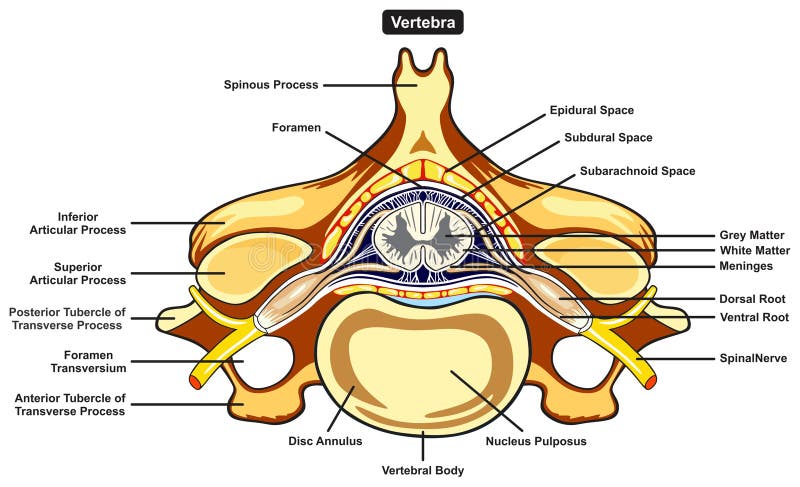
Anatomy Pathways - Spinal Cord and Spinal Nerves The dura mater ("tough mother") forms a sac from the foramen magnum where it is continuous with the brain dura mater. The dura is the most superficial of the three meninges that surround and protect both the brain and the spinal cord. The space superficial to the dura mater between it and the vertebral canal wall is the epidural space.

Spinal Cord Meninges Diagram | Quizlet
Anatomy of the spinal cord - e-Anatomy - IMAIOS The second illustration of the spinal cord schematised in 3-dimensions the white matter cords, the grey columns and the horns of the spinal cord. Next, the user will find anatomical sections of the spinal cord at different levels: cervical spinal cord (C2, C5), thoracic spinal cord (T10), lumbar spinal cord (L3) and sacral spinal cord (S3).

Frontiers | Imaging Ischemic and Hemorrhagic Disease of the ...
Spinal Cord: Function, Anatomy and Structure - Cleveland Clinic The spinal cord is a long, tube-like band of tissue. It connects your brain to your lower back. Your spinal cord carries nerve signals from your brain to your body and vice versa. These nerve signals help you feel sensations and move your body. Any damage to your spinal cord can affect your movement or function. Appointments 866.588.2264
Meninges - Wikipedia
Spinal Cord and Spinal Nerve Worksheet.docx - Spinal Cord... Label the 5 steps of a reflex: 2. The dorsal/posterior white matter of the spinal cord consists primarily of sensory and motor tracts. The columns of the white matter are called funiculi,the tracts within are called ascending and descending tracts. 3. Differentiate the structure and function of the white and grey matter of the spinal cord.
Spinal cord: Anatomy, functions, and injuries
Penetration of Drugs through the Blood ... - PubMed Central … WebInterrupted arrows indicate where a diffusion of water or solutes can occur between brain capillaries, CSF, and nervous tissue: (a) across the blood-brain barrier; (b) across the epithelium of the choroid plexus; (c) across the ependyma; (d) across the pia-glial membranes at the surface of the brain and spinal cord; (e and f) across the cell …
Spinal cord | Encyclopedia | Anatomy.app | Learn anatomy | 3D ...
› 40600784 › LIBRO_PARA_COLOREAR_NETTER(PDF) LIBRO PARA COLOREAR NETTER - Academia.edu Background: The aim of our study was to examine the effect of mild maternal hypothyroidism on the apoptosis of the oocytes in the ovaries of rats in the early postnatal period during formation of oocytes and follicles.
Chapter 13 Lecture Outline - ppt download
Spinal Cord Diagram with Detailed Illustrations and Clear Labels - BYJUS The spinal cord is one of the most important structures in the human body. It is the most important structure for any vertebrate. Anatomically, the spinal cord is made up of nervous tissue and is integrated into the spinal column of the backbone. Main Article: Spinal Cord - Anatomy, Structure, Function, and Spinal Cord Nerves; Also Read:

spinal cord | anatomy | Britannica
Spinal cord, Meninges and nerve connective tissues - Quiz 12 Questions Show answers. Q. The 3 layers of meninges from superficial to deep are. Q. Where is CSF located within the meninges? Q. Which number represents the gray matter. Q. Which number represents the white matter?

Pin on Brain Food
quizlet.com › 457717968 › chapter-14-assignmentChapter 14 Assignment Flashcards | Quizlet The meninges is a three-layered, membranous covering of the brain and spinal cord. Read the descriptions below and then click and drag them into the appropriate box based on whether they pertain to the dura mater, the arachnoid mater, or the pia mater. 1. Dura Mater 2. Arachnoid Mater 3. Pia Mater 1. The most superficial layer 2. The deepest ...
THE NERVOUS SYSTEM
Wikipedia, the free encyclopedia WebThe A and B Loop is a streetcar circle route of the Portland Streetcar system in Portland, Oregon, United States.Operated by Portland Streetcar, Inc. and TriMet, it consists of two services within the Central City that travel a loop between the east and west sides of the Willamette River by crossing the Broadway Bridge (pictured) in the north and Tilikum …

spinal meninges and structure of the spinal cord figure 16.2 ...
Correctly Label The Following Anatomical Features Of The Spinal Cord. Correctly label the following anatomical features of the cerebellum. 26 pimate dura materidura shout arachnoid mater meninges spinal cord farinebidural space derttelaments subdural cu ganglion 1 points posterior references meninges anterior (*) spinal cord and wertebra (cervical this is the most superficial.

The Spinal Cord, Spinal Nerves, and Spinal Reflexes - ppt ...
Solved Check my work 2 Label the spinal cord meninges and | Chegg.com Expert Answer. Answer is labeled in below image : • About : - The subarachnoid space is the interval between the arachnoid membrane and t …. View the full answer. Transcribed image text: Check my work 2 Label the spinal cord meninges and spaces. 1 Subarachnoid space Subdural space Dura mater Pia mater Arachnoid mater points eBook Print ...

Lab Quiz #1 (no tracts) Flashcards | Quizlet
Anatomical Body Planes | Science Trends WebThe dorsal cavity is one long continuous cavity that houses portions of the central nervous system including the spinal cord and brain. It is found on the body’s dorsal side. The cranial cavity contains the cerebrospinal fluid, the brain’s meninges, and the brain itself. The cranial cavity overlaps with the anterior portion of the dorsal cavity. The ventral cavity is where …

The Spinal Cord in Hindi - YouTube
› brain_tumor › articleBrain Tumors: Types, Causes, 13 Warning Signs, Treatment ... Apr 27, 2022 · Spinal tap: Your doctor may remove a sample of cerebrospinal fluid (the fluid that fills the spaces in and around the brain and spinal cord). This procedure is performed with local anesthesia. The doctor uses a long, thin needle to remove fluid from the lower part of the spinal column. A spinal tap takes about 30 minutes.

Neuraxial Anatomy - NYSORA | NYSORA
Spinal Cord Practice Material Answers.pdf - 1. List the key spaces and ... List the key spaces and meninges surrounding the spinal cord from outermost to innermost. Epidural space, dura mater, subdural space, arachnoid mater, subarachnoid space, pia mater Epidural space , dura mater , subdural space , arachnoid mater , subarachnoid space , pia mater 2.

Solved Conus Medoll 13 lumbar enlargement carda eguna Filum ...
Spinal Meninges Anatomy, Diagram & Function | Body Maps - Healthline Pia mater: The innermost layer, the pia mater hugs the spinal cord and brain like a coat. It has blood vessels that deliver oxygen and nutrients to the spinal cord. To check for problems of the CNS...
Spinal Meninges
Meninges Cerebrales: Layers and Spaces (with Images) It is also called the leptomeningeal space, and is a fine space that exists between the arachnoid membrane and the pia mater. It contains arachnoid cords, as well as nerve and vascular structures. There are certain places where these spaces are wider and communicate with each other, called subarachnoid cisterns.

Spinal Cord Meninges Model (Transverse) Diagram | Quizlet
› pmc › articlesPenetration of Drugs through the Blood-Cerebrospinal Fluid ... The intracranial space and vertebral canal cannot be looked at as a single physiological compartment: it is divided into the CSF space and the extracellular and intracellular spaces of the brain and spinal cord (Fig. (Fig.1) 1) (42, 43). Within the individual parts of the same compartment, concentrations often differ . After i.v. injection ...
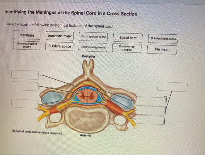
Solved Identifying the Meninges of the Spinal Cord in a ...
Arachnoid granulation - Wikipedia WebArachnoid granulations (also arachnoid villi, and pacchionian granulations or bodies) are small protrusions of the arachnoid mater (the thin second layer covering the brain) into the outer membrane of the dura mater (the thick outer layer). They protrude into the dural venous sinuses of the brain, and allow cerebrospinal fluid (CSF) to exit the subarachnoid …
The Spinal Cord, Spinal Nerves, and Spinal Reflexes
Meninges: What They Are & Function - Cleveland Clinic There are three spaces within the meninges: The epidural space is a space between your skull and dura mater and the dura mater of your spinal cord and the bones of your vertebral column. Analgesics (pain medicine) and anesthesia are sometimes injected into this space along your spine.
Spinal cord: Anatomy, structure, tracts and function | Kenhub
Spinal Cord - Internal Structure and Meninges - AnatomyZone You can see in this diagram the black line represents the dura mater, so this goes round the outside of the spinal cord and the structures that are associated with it. And then the light blue line represents the arachnoid mater, so between the dura mater and the arachnoid mater, you've got this space called the subdural space.
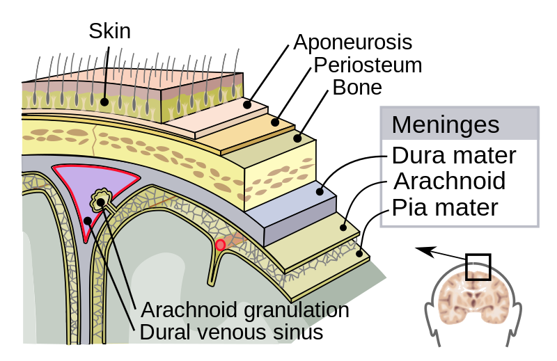
Dura mater - Wikipedia
Meninges Layers, Function & Anatomy - Study.com The brain and spinal cord are important structures that control the nervous system. These organs are covered by layers of tissues called meninges. There are three layers of meninges: Dura Mater...

14.2 Blood Flow the meninges and Cerebrospinal Fluid ...
The Meninges - Dura - Arachnoid - Pia - TeachMeAnatomy The meninges refer to the membranous coverings of the brain and spinal cord. There are three layers of meninges, known as the dura mater, arachnoid mater and pia mater. These coverings have two major functions: Provide a supportive framework for the cerebral and cranial vasculature.
Spinal cord: Anatomy, structure, tracts and function | Kenhub
› image › nervovNervous System: Explore the Nerves with Interactive Anatomy ... Nov 02, 2020 · Each spinal nerve exits from the spinal cord through the intervertebral foramen between a pair of vertebrae or between the C1 vertebra and the occipital bone of the skull. Meninges The meninges are the protective coverings of the central nervous system (CNS).
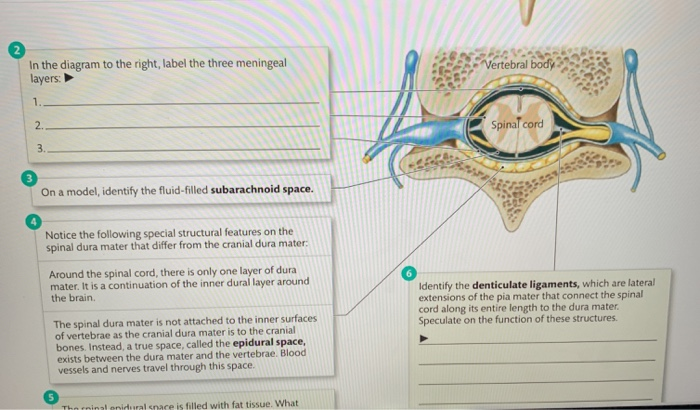
Solved In the diagram to the right, label the three | Chegg.com

The brain and spinal cord | Canadian Cancer Society
Body Cavities Labeled: Organs, Membranes, Definitions ...

A&P 17 Lab.pdf - 11/22/17, 11:00 AM PRINTED BY: ss3547@mynsu ...
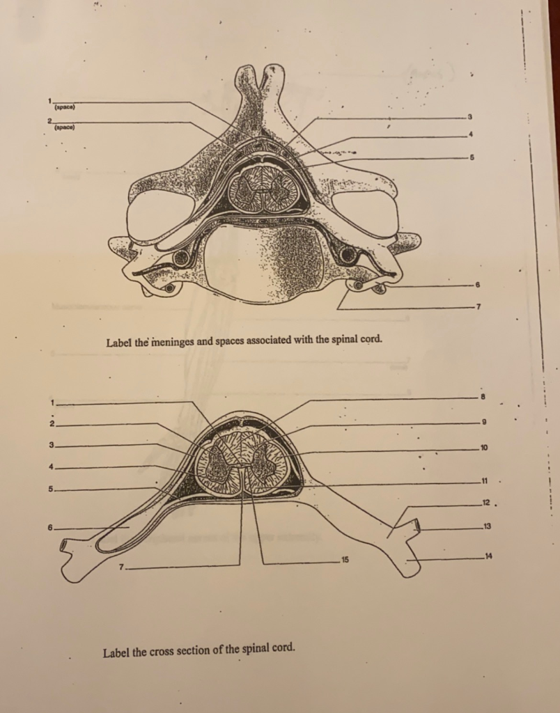
Solved (space) 2 (space) Label the meninges and spaces ...
Meninges Stock Illustrations – 226 Meninges Stock ...

SPINAL CORD WEEK 8 Flashcards | Quizlet
Meninges: Dura, arachnoid, pia, meningeal spaces | Kenhub

Introduction to Meningitis - Brain, Spinal Cord, and Nerve ...
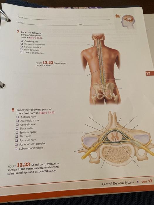
Solved Section 7 Label the following parts of the spinal ...
0 Response to "40 label the spinal cord meninges and spaces."
Post a Comment