40 picture of microscope with label
Microscope picture label Flashcards | Quizlet Start studying Microscope picture label. Learn vocabulary, terms, and more with flashcards, games, and other study tools. Guide to Teaching Kids About Cells | Science Explorers 25-04-2019 · Give each student a chance to look through the microscope at the cells. Point out that each slide contains numerous cells. Repeat the process with the second slide. Have students draw pictures of what they saw under the microscope and guess what the cells do. Finish with an explanation of the cell and its organelle functions.
Microscope Parts, Function, & Labeled Diagram - slidingmotion Microscope Parts Labeled Diagram The principle of the Microscope gives you an exact reason to use it. It works on the 3 principles. Magnification Resolving Power Numerical Aperture. Parts of Microscope Head Base Arm Eyepiece Lens Eyepiece Tube Objective Lenses Nose Piece Adjustment Knobs Stage Aperture Microscopic Illuminator Condenser Lens

Picture of microscope with label
Parts of a Simple Microscope - Labeled (with diagrams) A simple microscope is a very first type of microscope ever created. It consists of simple parts and performs simple functions. Although there are now many advanced microscope types, some applications may still demand the use of a simple microscope. In this article, we are going to discuss the parts and functions of a simple microscope. Microscopes | Biomedx Microscope System Limited Warranty Biomedx warrants that the microscope systems it sells and any related accessories bearing the Biomedx label (individually a “Product” and collectively the “Products”) will be free from defects in materials and workmanship under normal use and service for a period, beginning from the date of purchase, of ; Lungs (Human Anatomy): Picture, Function, Definition, Conditions - WebMD WebMD's Lungs Anatomy Page provides a detailed image and definition of the lungs. Learn about lung function, problems, location in the body, and more.
Picture of microscope with label. Drawing Of A Microscope And Label - Warehouse of Ideas Yet even with the technology to digital capture images, many scientists still depend on their abilities to sketch microscope slides. Drawn as seen through 400x magnification). Here presented 54+ microscope drawing and label images for free to download, print or share. Title Is Informative, Centered, And Larger Than Other Text. Parts of a Microscope Labeling Activity - Storyboard That In this activity, students will create a poster of a microscope with labeled parts. Students will identify and describe the microscope parts and functions. This is an awesome activity to complete at the beginning of either the school year or the unit on basic cells. ... All storyboards and images are private and secure. Teachers can view all of ... Cell Size and Scale - University of Utah Smaller cells are easily visible under a light microscope. It's even possible to make out structures within the cell, such as the nucleus, mitochondria and chloroplasts. Light microscopes use a system of lenses to magnify an image. The power of a light microscope is limited by the wavelength of visible light, which is about 500 nm. Microscopes | Biomedx Microscope System Limited Warranty Biomedx warrants that the microscope systems it sells and any related accessories bearing the Biomedx label (individually a “Product” and collectively the “Products”) will be free from defects in materials and workmanship under normal use and service for a period, beginning from the date of purchase, of ;
Simple Microscope - Diagram (Parts labelled), Principle, Formula and Uses The working principle of a simple microscope is that when a lens is held close to the eye, a virtual, magnified and erect image of a specimen is formed at the least possible distance from which a human eye can discern objects clearly. Magnification formula The magnification power of a simple microscope is expressed as: M = 1 + D/F Where Microscope Labeled Pictures, Images and Stock Photos Microscope Labeled Pictures, Images and Stock Photos View microscope labeled videos Browse 49 microscope labeled stock photos and images available, or start a new search to explore more stock photos and images. Newest results Fluorescent Imaging immunofluorescence of cancer cells growing... Microscope diagram vector illustration. Guide to Teaching Kids About Cells | Science Explorers Apr 25, 2019 · Give each student a chance to look through the microscope at the cells. Point out that each slide contains numerous cells. Repeat the process with the second slide. Have students draw pictures of what they saw under the microscope and guess what the cells do. Finish with an explanation of the cell and its organelle functions. Labeling the Parts of the Microscope | Microscope World Resources Labeling the Parts of the Microscope This activity has been designed for use in homes and schools. Each microscope layout (both blank and the version with answers) are available as PDF downloads. You can view a more in-depth review of each part of the microscope here. Download the Label the Parts of the Microscope PDF printable version here.
Label Microscope Diagram - EnchantedLearning.com Using the terms listed below, label the microscope diagram. arm - this attaches the eyepiece and body tube to the base. base - this supports the microscope. body tube - the tube that supports the eyepiece. coarse focus adjustment - a knob that makes large adjustments to the focus. diaphragm - an adjustable opening under the stage, allowing ... Microscope With Labels clip art | Microscope parts, Scientific method ... Microscope With Labels clip art | Microscope parts, Scientific method, Science diagrams From clker.com vector clip art online, royalty free & public domain Download Clker's Microscope With Labels clip art and related images now. Multiple sizes and related images are all free on Clker.com. D Dixie Tsutsaeva 2k followers More information Microscopy - Wikipedia The field of microscopy (optical microscopy) dates back to at least the 17th-century.Earlier microscopes, single lens magnifying glasses with limited magnification, date at least as far back as the wide spread use of lenses in eyeglasses in the 13th century but more advanced compound microscopes first appeared in Europe around 1620 The earliest practitioners of microscopy include Galileo ... Microscope Parts and Functions Most specimens are mounted on slides, flat rectangles of thin glass. The specimen is placed on the glass and a cover slip is placed over the specimen. This allows the slide to be easily inserted or removed from the microscope. It also allows the specimen to be labeled, transported, and stored without damage.
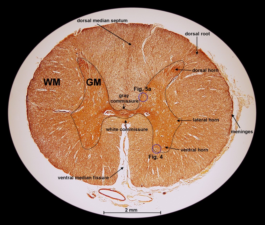
Exploration of the Human Spinal Cord
Microscopy - Wikipedia Optical or light microscopy involves passing visible light transmitted through or reflected from the sample through a single lens or multiple lenses to allow a magnified view of the sample. The resulting image can be detected directly by the eye, imaged on a photographic plate, or captured digitally.The single lens with its attachments, or the system of lenses and imaging equipment, …

100pcs/box biological microscope specimens Prepared Glass Microscope ...
XGT-9000 - HORIBA In this application note, we carried out elemental map imaging on edible crickets using a HORIBA XGT-9000 X-ray analytical microscope and revealed the rich source of zinc in their jaws. Read more Foreign Matter Analysis in Food using the XGT 9000
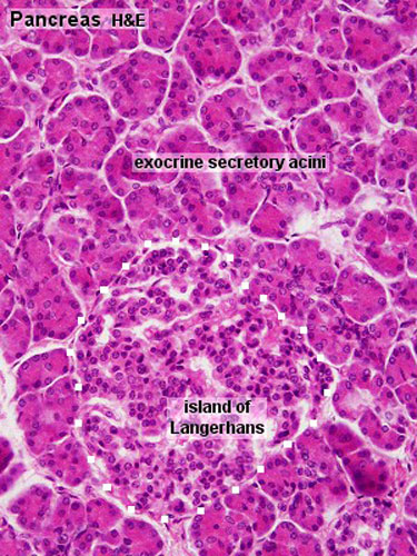
Gastrointestinal Tract - Pancreas Histology - Embryology
Virtual Labs: Using the Microscope - GameUp - BrainPOP. In this free online science interactive, students learn the procedures for operating a compound optical light microscope as they would use in a science lab. bVX0-zncj9qJ3G1_r18rkIpQL02X-Oi6tWViR4g4-vwDVmU50WZA-4bRZMjM2TXmc88PAkJ1g0jIembnEbM
Search in gallery
PDF Parts of a Microscope Printables - Homeschool Creations Label the parts of the microscope. You can use the word bank below to fill in the blanks or cut and paste the words at the bottom. Microscope Created by Jolanthe @ HomeschoolCreations.net. Parts of a eyepiece arm stageclips nosepiece focusing knobs illuminator stage objective lenses

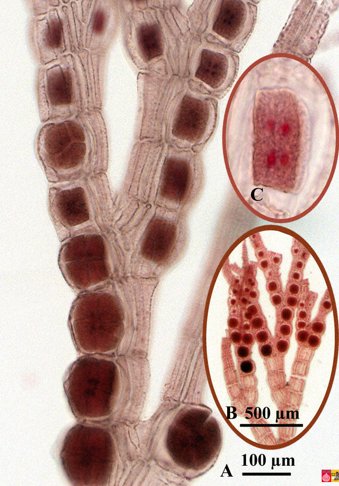
UBC Biology 210 Blog: Lab 5 - Leaves and Modifications
UD Virtual Compound Microscope - University of Delaware ©University of Delaware. This work is licensed under a Creative Commons Attribution-NonCommercial-NoDerivs 2.5 License.Creative Commons Attribution-NonCommercial-NoDerivs 2
Print Structure and Function of Plants (Lab Practical #1) flashcards ...
PDF Label parts of the Microscope Label parts of the Microscope: . Created Date: 20150715115425Z

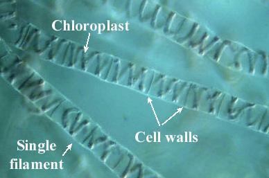
Under the Micrsocope: Onion Cell (100x - 400x) - YouTube
Microscope Labeling - The Biology Corner Students label the parts of the microscope in this photo of a basic laboratory light microscope. Can be used for practice or as a quiz. ... Microscope Labeling . Microscope Use: 15. When focusing a specimen, you should always start with the _____ objective. 16. When using the high power objective, only the _____ knob should be used. 17. The ...
0 Response to "40 picture of microscope with label"
Post a Comment