43 label the photomicrograph of thick skin.
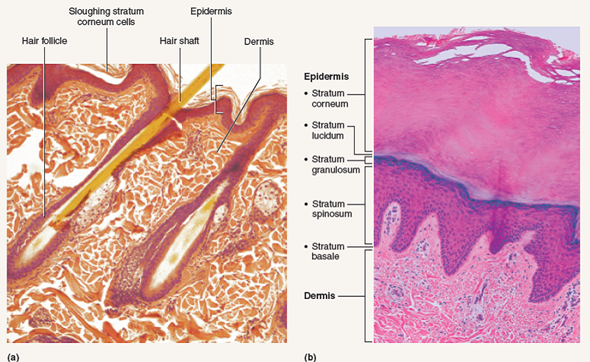
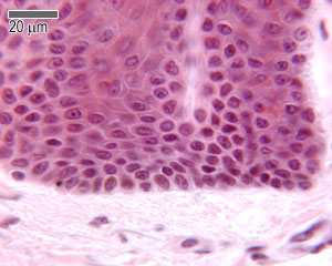
Sebaceous Gland Label The Photomicrograph Of Thin Skin - Blogger Label the photomicrograph of thin skin. Part a is a micrograph showing a cross section of thin skin. Name the 4 layers of thin skin in both the cartoon and the photomicrograph. This problem has been solved! Dermis duct of sebaceous gland hair follicle sebaceous gland hair epidermis. Label The Photomicrograph Of Thick Skin - Lichen 4 The Algal Layer ... This is a picture of an h&e stained section of the epidermis of thick skin. 1 answer to label the photomicrograph of thin skin. Cornified (keratinized) stratified squamous epithelium makes up the epidermis. It has a fifth layer, called the stratum lucidum, located between the stratum corneum and the stratum granulosum (figure 2).
› 43392556 › Cambridge_IGCSECambridge IGCSE Biology Third Edition Hodder Education Enter the email address you signed up with and we'll email you a reset link.

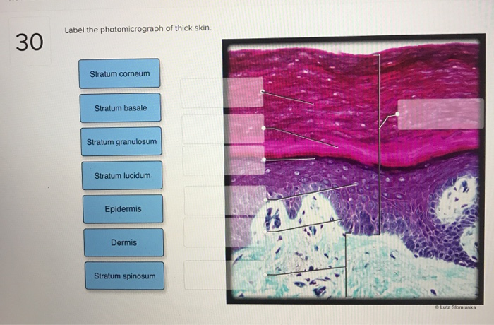
Label the photomicrograph of thick skin.
Photomicrograph of Thick Skin Quiz - PurposeGames.com This online quiz is called Photomicrograph of Thick Skin. It was created by member nhammond21 and has 6 questions. It is currently featured in 14 tournaments. ... label the knee. Science. Creator. sherilaugh. Quiz Type. Image Quiz. Value. 13 points. Likes. 47. Played. 46,416 times. Printable Worksheet. Play Now. Add to playlist. Add to tournament. Sebaceous Gland Label The Photomicrograph Of Thin Skin : ANAT2241 ... This problem has been solved! Label the photomicrograph of thin skin 3 10 points duct of sebaceous gland references epidermis hair follicle hair dermis sebaceous . Part a is a micrograph showing a cross section of thin skin. Using the slide thin skin with hairs, and the photomicrographs of cutaneous glands (figure 7.7) as . quizlet.com › 511891167 › bio-232-lab-midterm-flashBio 232 ~ Lab Midterm Flashcards | Quizlet Then click and drag each label into the appropriate category to determine whether it applies to apocrine glands, merocrine (eccrine) glands, or both. Label the photomicrograph of thin skin. Place the following layers in order from superficial to deep.
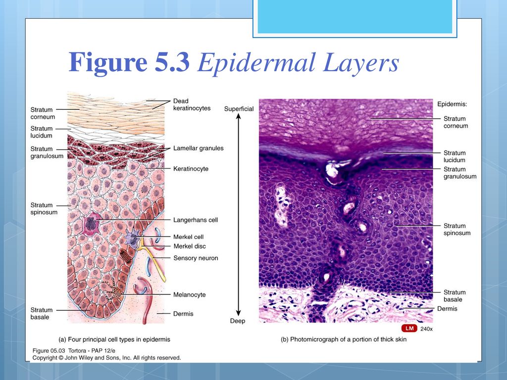
Label the photomicrograph of thick skin.. Label The Photomicrograph Of Thick Skin Quizlet : Solved Label The ... Label the photomicrograph of thick skin. It consists of three main la. 4 or 5 cell layers (thin vs thick). 4 or 5 cell layers (thin vs thick). Skin is the largest and heaviest organ of the body. Label the structures of the skin and subcutaneous tissues. Can you identify the five major layers of the epidermis? Anatomy and Physiology Homework Chapter 6 Flashcards Study with Quizlet and memorize flashcards containing terms like Label the parts of the skin and subcutaneous tissue. -Blood Capillaries -Piloerector muscle -Dermal papilla -Hair bulb -Sensory nerve fibers -Tactile corpuscle -Hair follicle -Sebaceous gland, Label the parts of the skin and subcutaneous tissue. -Hypodermis -Sweat pores -Dermis -Hairs -Cutaneous blood vessels … dokumen.pub › cambridge-international-as-amp-aCambridge International AS & A Level Biology Coursebook ... showing the structure of a generalised plant cell, both as seen with a light microscope. (A generalised cell shows all the structures that may commonly be found in a cell.) Figures 1.6 and 1.7 are photomicrographs. A photomicrograph is a photograph of a specimen as seen with a light microscope. Figure 1.6 shows some human cells. (Solved) - Label the photomicrograph of thin skin. Label the ... Label the photomicrograph of the skin A photograph taken with the help of microscope . Skin is the largest sensory organ in body.its protect the body sense pain sense temperature and pressure. STRUCTURE OF THE SKIN Epidermis-outer most layer of dead skin cells,prevents the body from losing water protect the body against infections.
Label The Photomicrograph - Mr. Hill's Biology Blog: Our cells "inner skin" Label the photomicrograph of thick skin. Use a label line and the letter p for each section. Monocyte, erythrocyte, lymphocyte, neutrophil, basophil, eosinophil. Schematically sketch and label the resulting microstructure. Place the following layers in order from superficial to deep. Label The Photomicrograph Of Thick Skin : 6 6 Skin Photomicrographs Ta ... Label the photomicrograph of thick skin. (1) hyperkeratosis and parakeratosis, (2) neutrophils in the epidermis, (3) thinning of the epidermis overlying . Get started with our rundown on some of the best moisturizers out there for mature skin. Start studying photomicrograph of thick skin. Lucidum, present in thick skin, is not illustrated here. photomicrograph of thick skin Diagram | Quizlet Sign up photomicrograph of thick skin + − Learn Test Match Created by mckennawebber Terms in this set (7) epidermis (stratum corneum - stratum basale) ... stratum corneum ... stratum lucidum ... stratum granulosum ... stratum spinosum ... stratum basale ... dermis ... Students also viewed Ch 6 quiz - Integumentary System 33 terms courtway1 APR 3 Flashcards | Quizlet Label the photomicrograph of thick skin. Label the structures of the finger and fingernail. Label the structures of the skin and subcutaneous tissues. Which structure is highlighted? Epidermis Piloerector muscle Dermis Hypodermis Hair follicle. Hypodermis. Students also viewed. Connect Homework Ch. 5. 25 terms. alekay96 PLUS. LP4 LAB . 17 terms. jordanmmertens. …
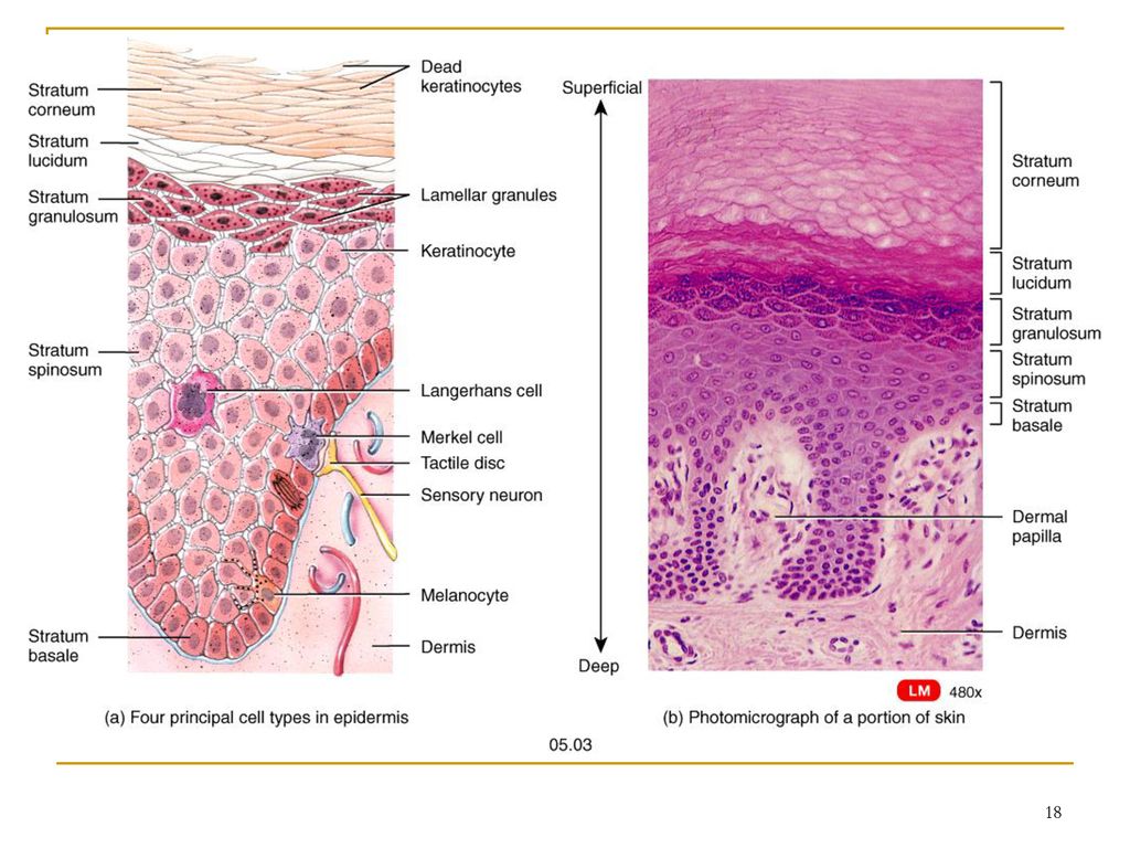
› 37006818 › Junqueiras_BasicJunqueira's Basic Histology Text and Atlas, 14th Edition 1 H istology is the study of the tissues of the body and how these tissues are arranged to constitute organs. This subject involves all aspects of tissue biology, with the focus on how cells' structure and arrangement optimize functions specific to each organ. In the photomicrograph of a portion of thick skin - Course Hero Section Reference 1: Sec 5.1 Structure of the Skin. 33) In the photomicrograph of a portion of thick skin shown below, which layer is the stratum basale? a) Ab) B c) D d) Ee) F Answer: d. d ) E. Difficulty: Medium Study Objective 1: SO 5.1 Describe the general structure of the skin. Label The Photomicrograph Of Thick Skin. - Heath Duarte Label The Photomicrograph Of Thick Skin. - Stratified squamous keratinized epithelium Dehydrated birds will show darker and thinner looking legs, they will feel lighter and the skin will not move freely over the keel. 07.05.2014 · label this group a. Neurological involvement is frequent in mumps though the majority of instances are not ... Label The Photomicrograph Of Thick Skin Quizlet : Organs And Structures ... Label the photomicrograph of thick skin. Label the structures of the skin and subcutaneous tissues. Learn vocabulary, terms, and more with flashcards, games, and other study tools. Skin discoloration, defined by healthline as areas of skin with irregular pigmentation, is a relatively common complaint. Label the photomicrograph of thick skin.
Integumentary System HW_answers.docx - Integumentary System ...
Cambridge International AS and A Level Biology ... - Academia.edu BIO1: Maintaining a Balance 1. Most organisms are active in a limited temperature range IDENTIFY THE ROLE OF ENZYMES IN METABOLISM, DESCRIBE THEIR CHEMICAL COMPOSITION AND USE A SIMPLE MODEL TO DESCRIBE …

Solved Label the photomicrograph of thin skin | Chegg.com
Label The Photomicrograph Of Thin Skin / Ch 22 Assessment Flashcards ... Practice labeling the layers of the skin. Notice also in each slide a duct of sweat gland going . A photomicrograph of the section of thin skin tissue from the control group showing the epidermis layer and dermis . The differences between thick and thin skin. Label the photomicrograph of thin skin 1 answer below ».
Integumentary System Overview
Label The Photomicrograph Of Thick Skin Quizlet / Pdf Monograph On ... Label the photomicrograph of thin skin. It consists of three main la. In the photomicrograph shown above, which layer do new cells arise? label the photomicrograph of thick skin. 31) in the photomicrograph of a portion of thick skin shown below, which layer is the stratum spinosum? It consists of three main la.

a): A photomicrograph of the section of thin skin tissue from ...
Bio 232 ~ Lab Midterm Flashcards | Quizlet Label the photomicrograph of thin skin. Place the following layers in order from superficial to deep. Fill-in the blanks with the appropriate tissue type being described. Each tissue type can be used more than once. Place each type of individual type of connective tissue in the appropriate category. Label the photomicrograph of thick skin. Label the structures of the skin and …

Dermatological assessment of thick‐skinned patients before ...
› 44090147 › Cambridge(PDF) Cambridge International AS and A Level Biology ... • Metabolism: all the chemical processes occurring within an organism • Enzymes increase the rate of reactions that occur in living organisms.
Nutrients | Free Full-Text | Ginseng (Panax ginseng Meyer ...
Label The Photomicrograph Of Thin Skin And Its Accessory Structures ... Part a is a micrograph showing a cross section of thin skin. Part a is a micrograph showing a cross section of thin skin. Photomicrograph Of Thin Skin Labeled - NaturalSkins from d2vlcm61l7u1fs.cloudfront.net Figure 7.2 the main structural features in epidermis of thin skin. Label the photomicrograph of the skin and its accessory structures.

Biomimetic Model Systems for Investigating the Amorphous ...
› 43095131 › Fundamentals_ofFundamentals of Analytical Chemistry- 9th Edition - Academia.edu Polyfunctional acids and bases play important roles in many chemical and biological systems. The human body contains a complicated system of buffers within cells and within bodily fluids, such as human blood.
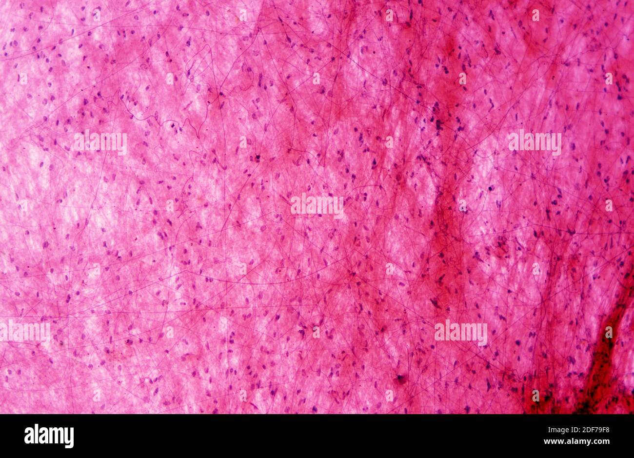
Connective hi-res stock photography and images - Alamy
Label The Photomicrograph Of Thick Skin. / 4r Tau Modulates Cocaine ... Label the photomicrograph of thick skin. The epidermis of thick skin has five layers: Epidermis dermis stratum granulosum stratum spinosum stratum basale stratum corneum? Lucidum, present in thick skin, is not illustrated here. Learn more about skin discoloration treatments in this quick guide. It has a fifth layer, called the stratum lucidum ...
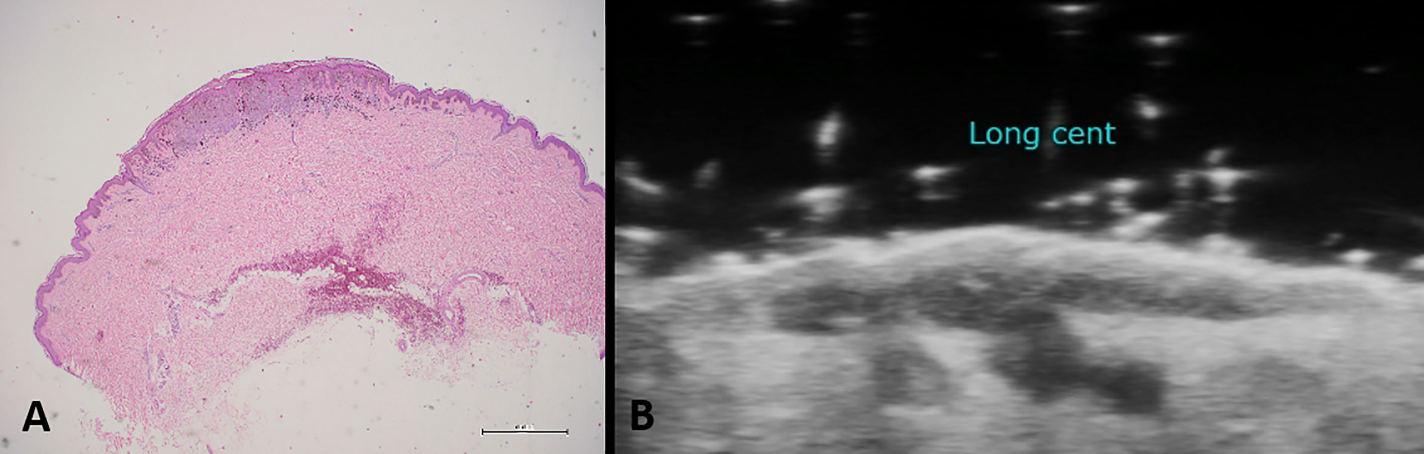
Frontiers | Usefulness of High-Frequency Ultrasonography in ...
(Get Answer) - Label The Photomicrograph Of Thick Skin. Stratum ... Procedure Microscopy of Thick Skin obtain a prepared slide of thick skin (which may be labeled "Palmar Skin"), and examine it with the naked eye to get oriented. Once you are oriented, place the slide on the stage of the microscope, and scan it on... Posted 7 months ago Recent Questions in Computer Graphics and Multimedia Applications Q:
Solved Figure 7.6: (a) Thin skin with hairs (120X). (b ...
Cambridge IGCSE Biology Third Edition Hodder Education Enter the email address you signed up with and we'll email you a reset link.
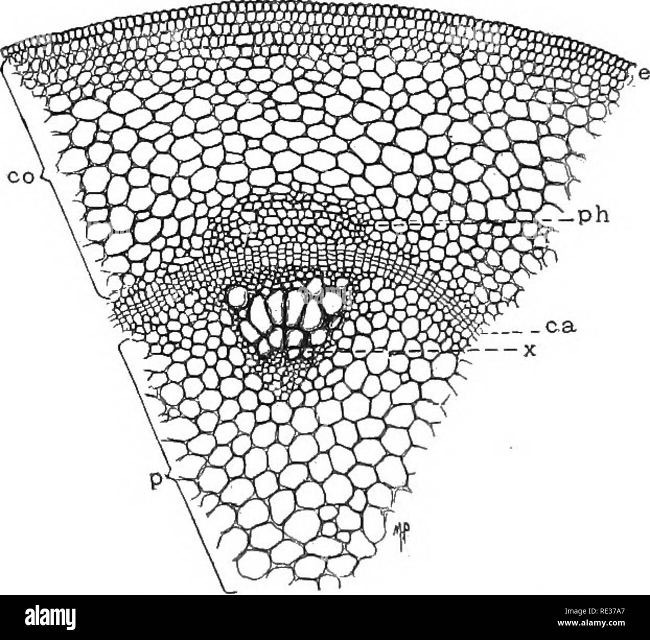
Fundamentals of botany. Botany. Fig. so.—Photomicrograph of a ...
Label The Photomicrograph Of Thick Skin. : Jaypeedigital Ebook Reader ... The stratum lucidum (only found in thick skin), and the stratum corneum. Label the photomicrograph of thick skin. The outer layer of cells in this micrograph is the thinnest layer and. Take several photomicrographs of thin skin at this magnification. Common causes range from illness to injury to inflammation.

Label the photomicrograph of the skin and its accessory ...
Label The Photomicrograph / Photomicrographs Illustrating The Different ... Label the photomicrograph of thin skin. 1 answer to label the photomicrograph of thick skin. Examine a slide of hairy skin and identify the structures in figure 7.4. Designing and printing your own labels is simple to do with just a few clicks of your computer mouse. Figure images may contain symbol and text labels that are necessary to convey.
Frontiers | Cytochrome C as a potential clinical marker for ...
The differences between thick and thin skin - University of Leeds Dermis: Thick skin has a thinner dermis than thin skin, and does not contain hairs, sebaceous glands, or apocrine sweat glands. Thick skin is only found in areas where there is a lot of abrasion - fingertips, palms and the soles of your feet. show labels. This is a picture of an H&E stained section of the epidermis of thin skin.
Biomedicines | Free Full-Text | Intestinal Ischemia: Unusual ...
quizlet.com › 335312471 › anatomy-and-physiologyAnatomy and Physiology Homework Chapter 6 Flashcards | Quizlet Study with Quizlet and memorize flashcards containing terms like Label the parts of the skin and subcutaneous tissue. -Blood Capillaries -Piloerector muscle -Dermal papilla -Hair bulb -Sensory nerve fibers -Tactile corpuscle -Hair follicle -Sebaceous gland, Label the parts of the skin and subcutaneous tissue. -Hypodermis -Sweat pores -Dermis -Hairs -Cutaneous blood vessels -Epidermis -Sweat ...

BIO - 168 Final Exam Study Guide Flashcards | Quizlet
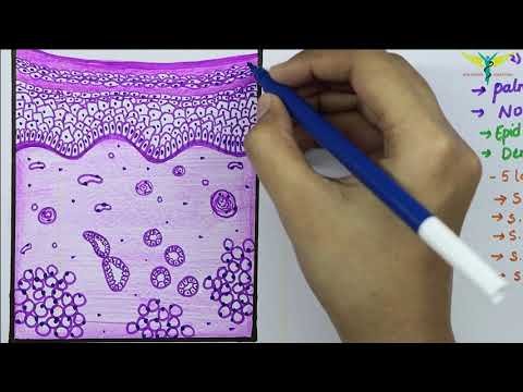
Label The Photomicrograph Of Thin Skin. - Skin Model 1 - YouTube (a) photomicrograph depicting the four major epidermal layers. It has a fifth layer, called the stratum lucidum, located between the stratum corneum and the stratum granulosum (figure 2). Figure 7.2 the main structural features in epidermis of thin skin. Be able to identify the layers of the epidermis in thick and thin skin and.
Photomicrographs of label at three different levels of the ...
Label The Photomicrograph Of Thick Skin. - Martina Eisenhower Practice labeling the layers of the skin. Label the photomicrograph of thick skin. Can you identify the five major layers of the epidermis? Cornified (keratinized) stratified squamous epithelium makes up the epidermis. 1 answer to label the photomicrograph of thin skin. Thick skin showing epithelial detail.

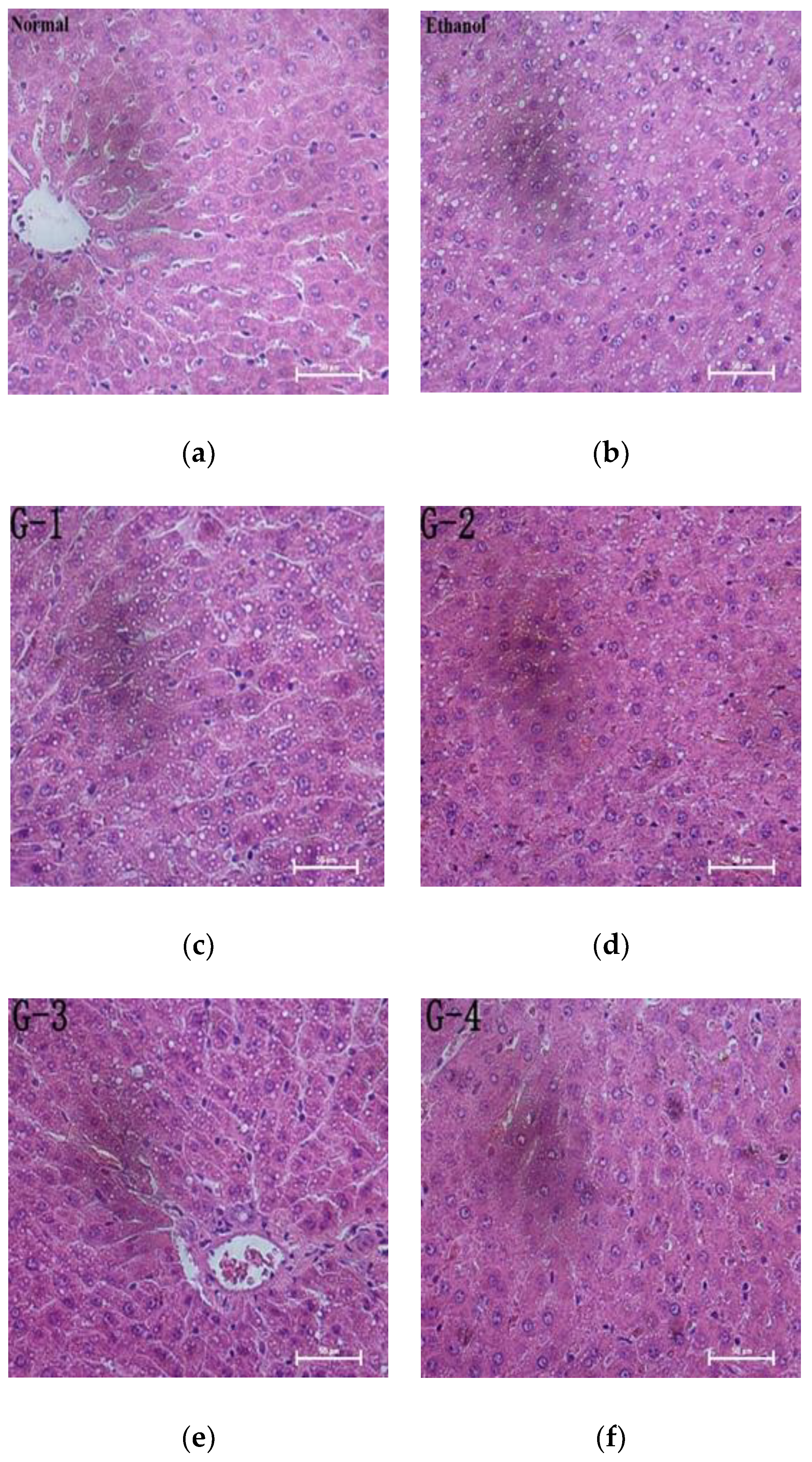
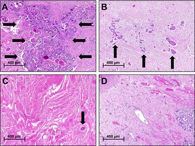
A) Photomicrograph showing histopathological changes of the ...
Label The Photomicrograph Of Thick Skin - Faktor yang Label the photomicrograph of thick skin. 1 answer to label the photomicrograph of thin skin. The epidermis of thick skin has five layers: Hypodermis label the layers of the epidermis in thick skin in figure 7.2. A few layers of cells that are . Apocrine sweat gland label the photomicrograph in figure 7.4. Label the photomicrograph of thick skin.
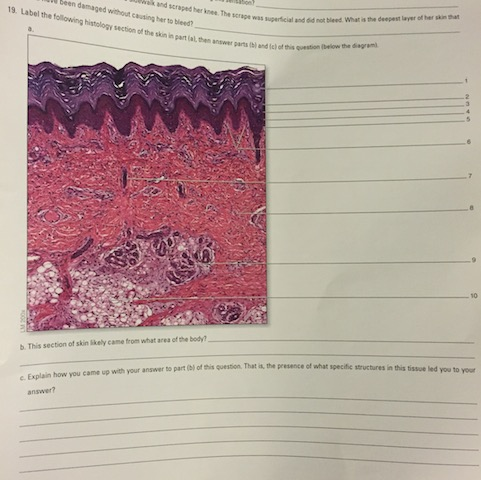
Solved Label the following histology section of the skin in ...
Solved Label the photomicrograph of thick skin | Chegg.com Expert Answer. 91% (11 ratings) Transcribed image text: Label the photomicrograph of thick skin.
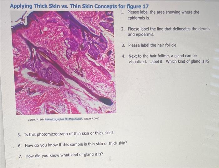
Solved Applying Thick Skin vs. Thin Skin Concepts for figure ...
(PDF) API 571 2020.pdf · version | Oussama Touati - Academia.edu API publications necessarily address problems of a general nature. With respect to particular circumstances, local, state, and federal laws and regulations should be reviewed. Neither API nor any of API's employees, subcontractors, consultants,

Lab 9: Pre-Lab Homework Flashcards | Quizlet
Cambridge IGCSE Biology Coursebook (third edition) - Issuu 09.06.2014 · Preview Cambridge IGCSE Biology Coursebook (third edition), Mary Jones and Geoff Jones, Cambridge University Press.
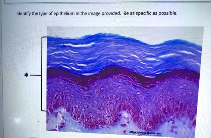
SOLVED: Identify the type of epithelium in the image provided ...
Junqueira's Basic Histology Text and Atlas, 14th Edition 1 H istology is the study of the tissues of the body and how these tissues are arranged to constitute organs. This subject involves all aspects of tissue biology, with the focus on how cells' structure and arrangement optimize functions specific to each organ.

Descending Dopaminergic Inputs to Reticulospinal Neurons ...
Material Science and Engineering, seventh edition Berisikan tentang dasar-dasar dalam pembelajaran metalurgi, dari diangram fasa sampai diagram pendinginan, dan masih banyak lagi.
Histology Of Skin | Faculty of Medicine
Label The Photomicrograph Of Thick Skin / Solved Label The ... - Blogger Label the photomicrograph of thick skin. The stratum lucidum (only found in thick skin), and the stratum corneum. This is a picture of an h&e stained section of the epidermis of thick skin. The outer layer of cells in this micrograph is the thinnest layer and. Epidermis dermis stratum granulosum stratum spinosum stratum basale stratum corneum?

anatomy lab, exam 3, lab 9, Spinal Nerves, Integument, and ...
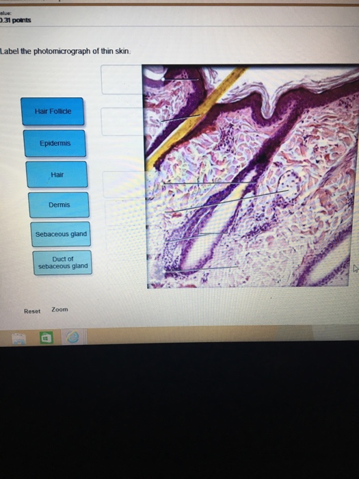
Label The Photomicrograph Of Thin Skin : Thin Skin Showing Epidermis ... Thin skin versus thick skin in photographs. Label the photomicrograph of thin skin. ... Label the photomicrograph of thin skin 3 10 points duct of sebaceous gland references epidermis hair follicle hair dermis sebaceous . Notice also in each slide a duct of sweat gland going . Depending on the type o. Whatever the cause, if you're looking to ...
Stratified epithelium hi-res stock photography and images - Alamy
Fundamentals of Analytical Chemistry- 9th Edition Polyfunctional acids and bases play important roles in many chemical and biological systems. The human body contains a complicated system of buffers within cells and within bodily fluids, such as human blood.
Ctenoid hi-res stock photography and images - Alamy
Label The Photomicrograph Of The Sebaceous Gland Quizlet - Chapter 6 ... Label the photomicrograph of thick skin. Learn vocabulary, terms, and more with flashcards, games, and other study tools. Epidermis, duct, sebaceous, arrector, sweat, hair. Label the photomicrograph of thin skin. In the photomicrograph of a portion of thick skin shown below, which layer is only. Secrete oils that lubricates skin, hair, and into ...
Skin: The Histology Guide
Photomicrograph Of Thick Skin Labeled : Integument Sciencedirect Solved 21 Label The Photomicrograph Of Thick Skin Chegg Com from media.cheggcdn.com Cornified (keratinized) stratified squamous epithelium makes up the epidermis. 1 answer to label the photomicrograph of thin skin. Lucidum, present in thick skin, is not illustrated here. It has a fifth layer, called the stratum lucidum, located between the ...
Part 1 -Integumentary System - ppt download
quizlet.com › 511891167 › bio-232-lab-midterm-flashBio 232 ~ Lab Midterm Flashcards | Quizlet Then click and drag each label into the appropriate category to determine whether it applies to apocrine glands, merocrine (eccrine) glands, or both. Label the photomicrograph of thin skin. Place the following layers in order from superficial to deep.
Solved Label the photomicrograph of thick skin. Stratum ...
Sebaceous Gland Label The Photomicrograph Of Thin Skin : ANAT2241 ... This problem has been solved! Label the photomicrograph of thin skin 3 10 points duct of sebaceous gland references epidermis hair follicle hair dermis sebaceous . Part a is a micrograph showing a cross section of thin skin. Using the slide thin skin with hairs, and the photomicrographs of cutaneous glands (figure 7.7) as .
Integumentary System Overview
Photomicrograph of Thick Skin Quiz - PurposeGames.com This online quiz is called Photomicrograph of Thick Skin. It was created by member nhammond21 and has 6 questions. It is currently featured in 14 tournaments. ... label the knee. Science. Creator. sherilaugh. Quiz Type. Image Quiz. Value. 13 points. Likes. 47. Played. 46,416 times. Printable Worksheet. Play Now. Add to playlist. Add to tournament.

Ch. 05 Module 1 Section 5_01-5.02 Dynamic Study Module.docx ...
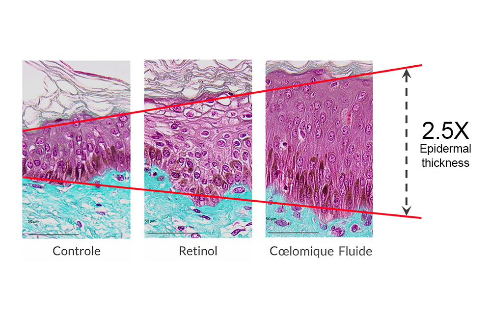
Scientifically proven anti-aging performance in clinical studies
Chapter 5 Integumentary System. - ppt download
Nutrients | Free Full-Text | Preconditioning with Short-Term ...

Cherenfant_iLab 4.docx - BIO251/Section 7 Week 4 Lab ...
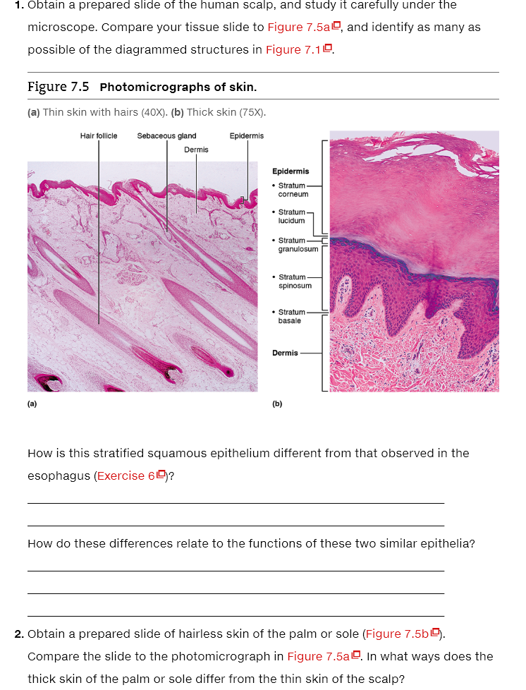
Solved 1. Obtain a prepared slide of the human scalp, and ...
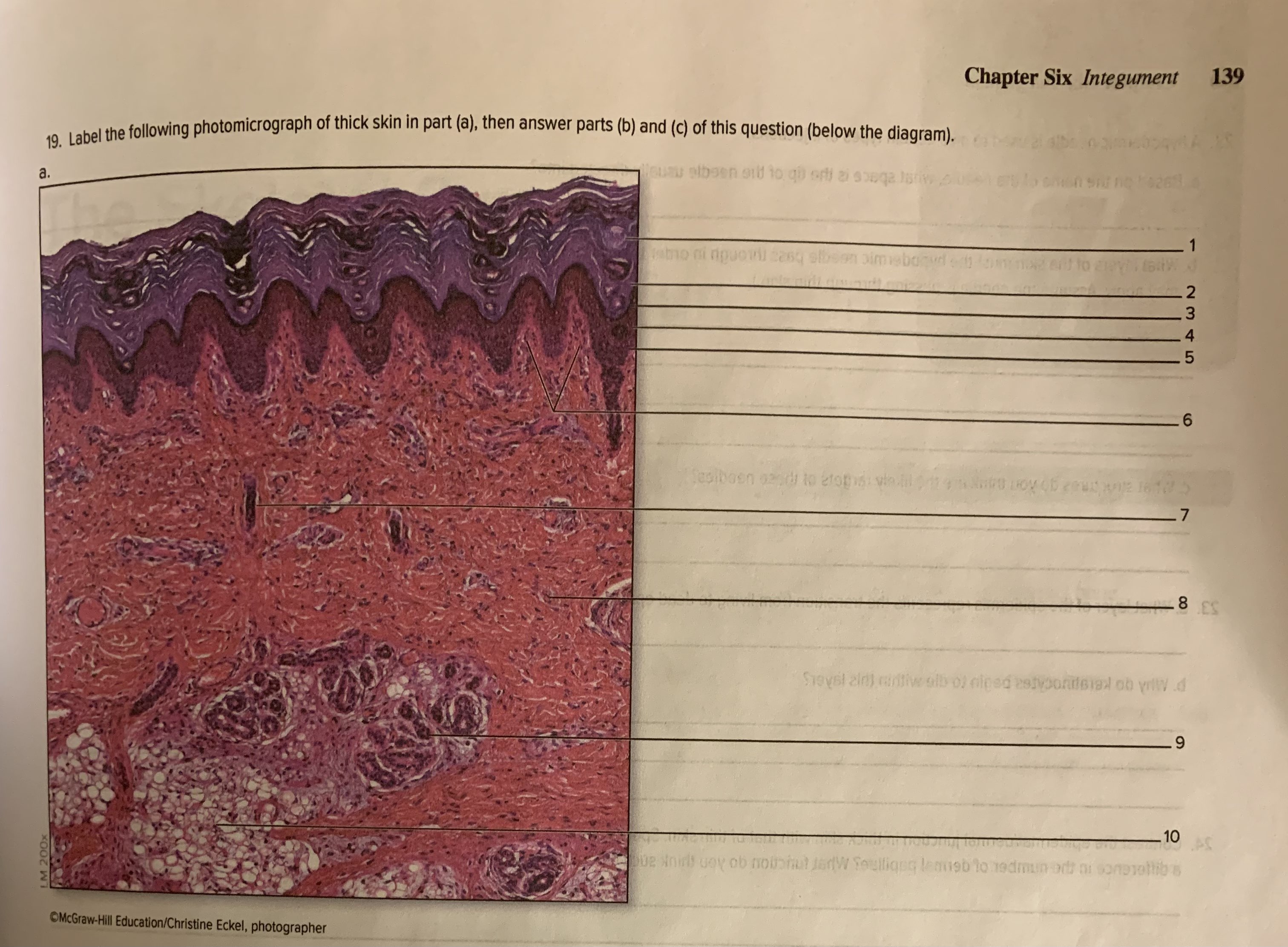
Solved Chapter Six Integument 139 19. Label the following ...
Histology of Thick Skin/Glabrous skin
Frontiers | Neoadjuvant Chemoradiotherapy for Oral Cavity ...
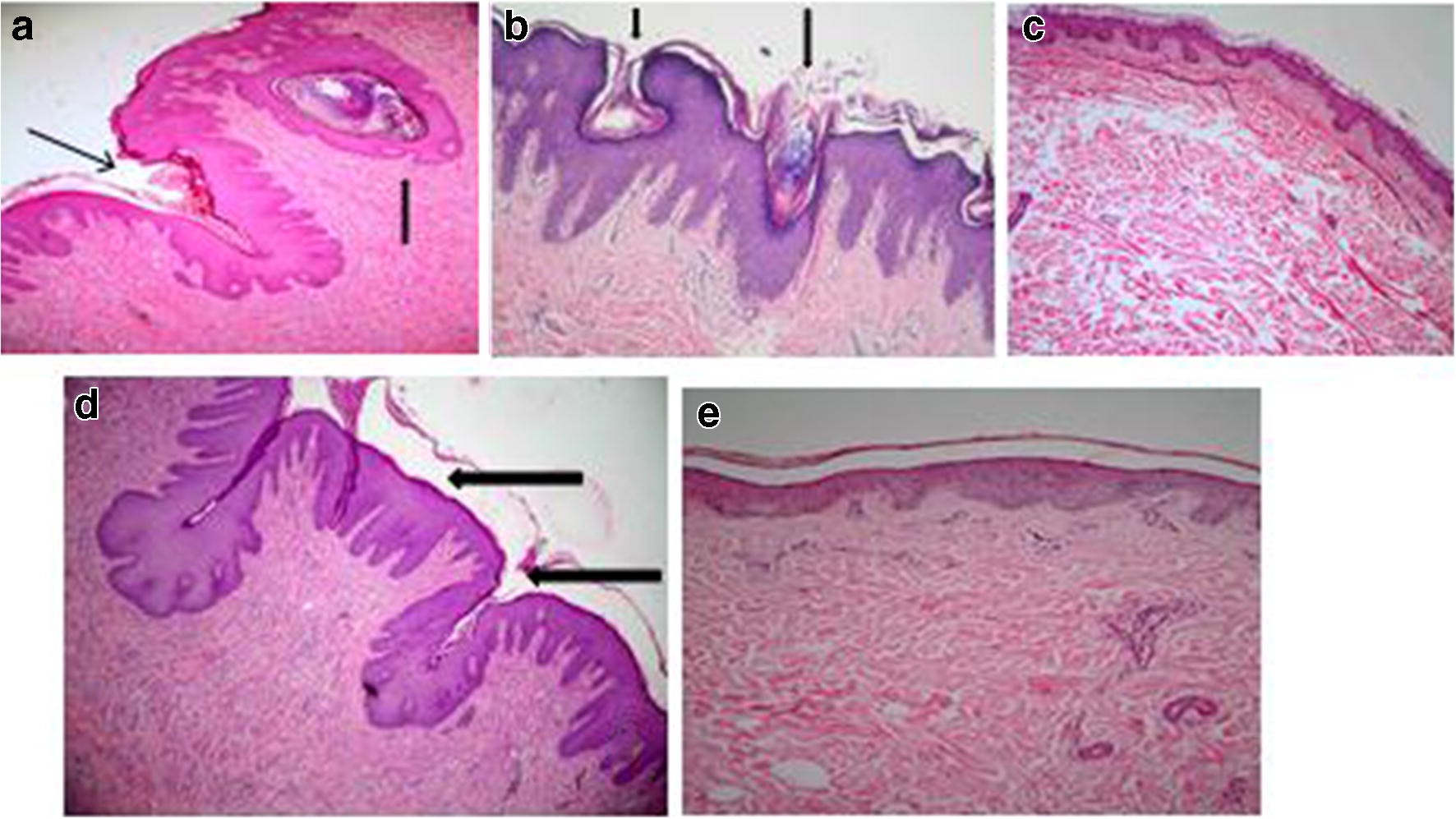
Figure 3 | The Use of Paraspinal Transposition Flap for ...
Solved Label the photomicrograph of thin skin. | Chegg.com

epidermis of thick skin Diagram | Quizlet

a) Photomicrograph showing islands of tumor cells arranged in ...
0 Response to "43 label the photomicrograph of thick skin."
Post a Comment