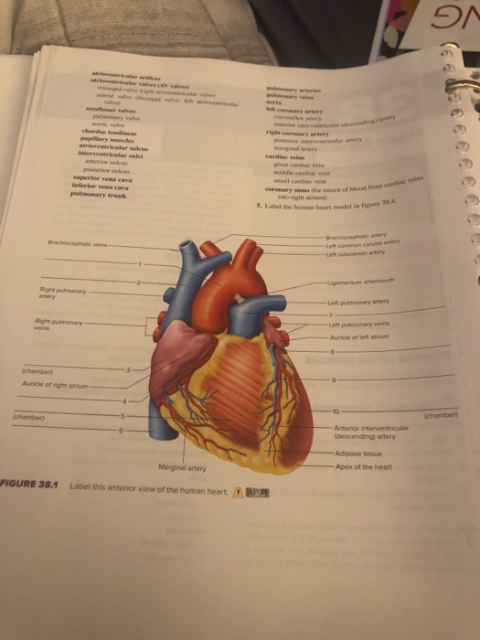
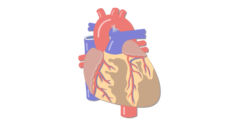
39 label this anterior view of the human heart
Heart Labeling anterior view Diagram | Quizlet Heart Labeling anterior view Diagram | Quizlet Heart Labeling anterior view 5.0 (2 reviews) + − Learn Test Match Created by Meghan12th Terms in this set (26) brachiocephalic trunk ... left common carotid artery ... superior vena cava ... aortic arch ... liigamentum arteriosum ... right pulmonary artery ... amending aorta ... right pulmonary veins Figure 34.1 label this anterior view of the human heart Figure 34.1 label this anterior view of the human heart. 24/11/2021 Client: muhammad11 Deadline: 2 Day. Laboratory. Manual for. Anatomy & Physiology. Connie Allen. Valerie Harper. 5e. Start Here, Go Anywhere. I dedicate this book to my new granddaughters, Gianna Leigh Madden and Taralyn Kay Thomas. —Connie Allen
Figure 34.1 label this anterior view of the human heart Figure 34.1 label this anterior view of the human heart. 06/01/2021 Client: saad24vbs Deadline: 14 Days. Laboratory. Manual for. Anatomy & Physiology. Connie Allen. Valerie Harper. 5e. Start Here, Go Anywhere. I dedicate this book to my new granddaughters, Gianna Leigh Madden and Taralyn Kay Thomas. —Connie Allen
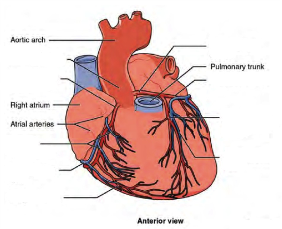
Label this anterior view of the human heart
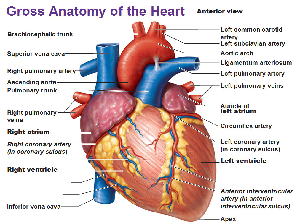
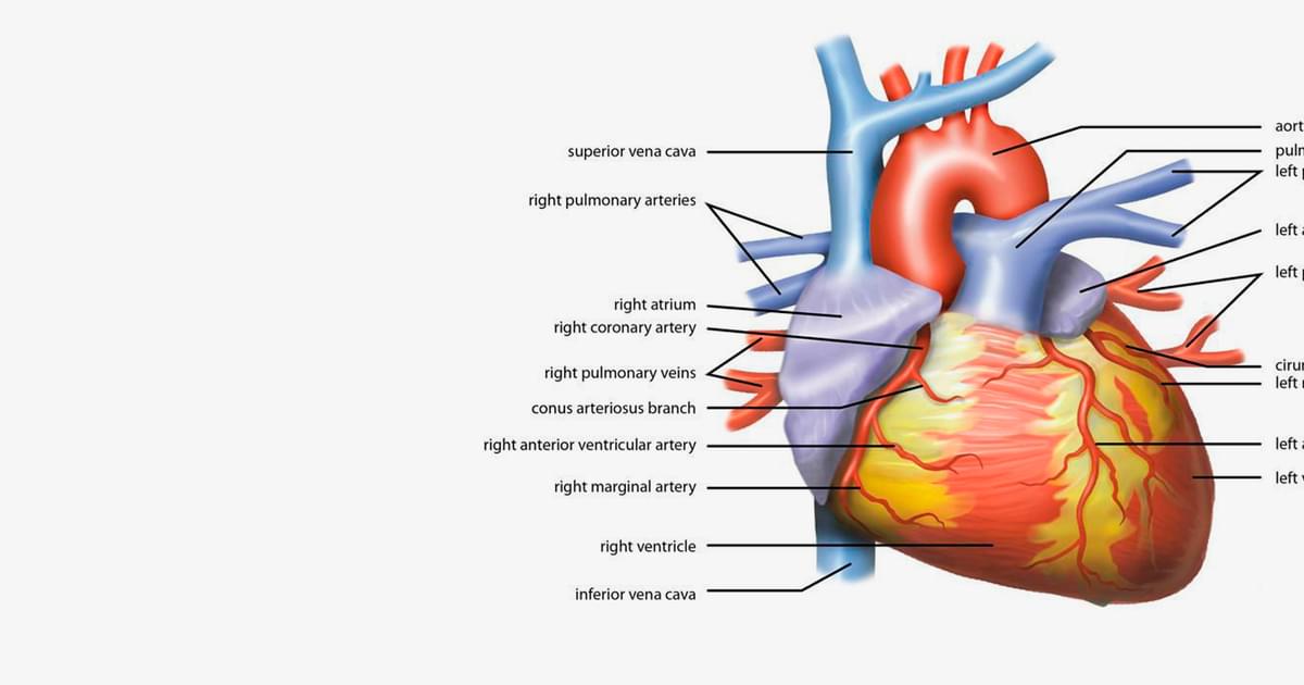
Human Heart - Diagram and Anatomy of the Heart - Innerbody The AV valve on the right side of the heart is called the tricuspid valve because it is made of three cusps (flaps) that separate to allow blood to pass through and connect to block regurgitation of blood. The AV valve on the left side of the heart is called the mitral valve or the bicuspid valve because it has two cusps. Gross Anatomy of Heart - Diagram Anterior View Flashcards - Quizlet Gross Anatomy of Heart - Diagram Anterior View Flashcards Learn Test Match Flashcards Learn Test Match Created by bk2811 MVHS HAP HEART Terms in this set (26) Brachiocephalic trunk 1 Superior vena cava 2 Right pulmonary artery 3 Ascending aorta 4 Pulmonary trunk 5 Right pulmonary veins 6 Right atrium 7 Right coronary artery (in coronary sulcus) 8 Anterior External View of Heart Labeling Diagram | Quizlet Anterior External View of Heart Labeling 5.0 (1 review) + − Learn Test Match Created by randajsmith Terms in this set (14) Superior Vena Cava ... Superior Vena Cava (SVC) ... Right Pulmonary Artery ... Right Pulmonary Veins ... Right Atrium ... Right Coronary Artery ... Right Ventricle ... Aorta ... Left pulmonary artery ... Left Atrium ...
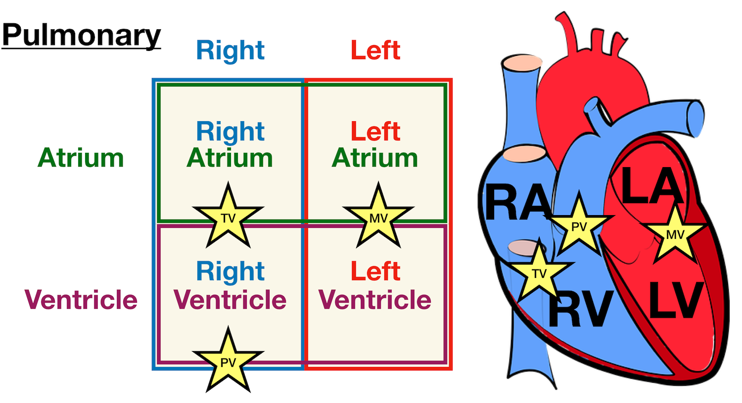
Label this anterior view of the human heart. 19.1 Heart Anatomy - Anatomy & Physiology The human heart is located within the thoracic cavity, medially between the lungs in the space known as the mediastinum. ... Figure 19.1.8 - Internal Structures of the Heart: This anterior view of the heart shows the four chambers, the major vessels and their early branches, as well as the four valves. The presence of the pulmonary trunk and ... Heart Anatomy | Anatomy and Physiology II | | Course Hero The human heart is located within the thoracic cavity, medially between the lungs in the space known as the mediastinum. Figure 1 shows the position of the heart within the thoracic cavity. ... Figure 8. This anterior view of the heart shows the four chambers, the major vessels and their early branches, as well as the valves. The presence of ... Anterior View of the Human Heart (preview) - YouTube If we were to strip everything you have on your chest from skin, to fat, and other tissues, and expose a little bit of the organs that you find within your chest or within the thorax, you would... Heart Anatomy: Labeled Diagram, Structures, Blood Flow ... - EZmed We now have a 2x2 table in which we can label the boxes/chambers of the heart. Box 1: The first box is located in the right upper region. We know the atria are on top, and since box 1 is located on the right side, this is the right atrium. Box 2: The second box is also located on the right side, but now we are in the lower region.
A Labeled Diagram of the Human Heart You Really Need to See The human heart, comprises four chambers: right atrium, left atrium, right ventricle and left ventricle. The two upper chambers are called the left and the right atria, and the two lower chambers are known as the left and the right ventricles. The two atria and ventricles are separated from each other by a muscle wall called 'septum'. External anterior heart labeling Quiz - PurposeGames.com This online quiz is called External anterior heart labeling. It was created by member stephanierotan and has 27 questions. ... Anatomy of the Human Heart - Internal Structures. Science. Creator. orkide1. Quiz Type. Image Quiz. Value. 24 points. Likes. 288. Played. ... External structure of the cerebral hemispheres view 1. by stephanierotan. 162 ... Heart chambers and associated great vessels - Anatomy Image: Anterior view of the human heart with labels. right atrium has two basic parts: a smooth posterior and an anterior portion in which bundles of muscle tissue form ridges in the walls. The muscle bundles are called pectinate muscles because they look like the teeth of a comb. Heart Diagram with Labels and Detailed Explanation - BYJUS The heart wall is made up of three layers: The outer layer of the heart wall is called epicardium. The middle layer of the heart wall is called myocardium. The inner layer of the heart wall is called endocardium. The heart consists of four valves: The aortic valve that prevents the backflow of blood when it is pumped from left ventricle to aorta.
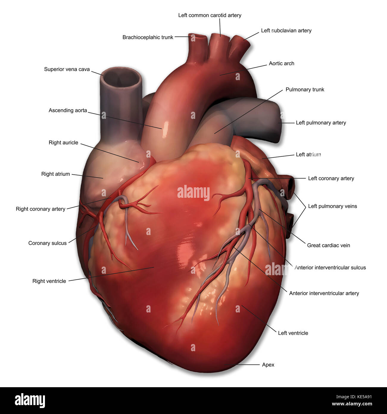
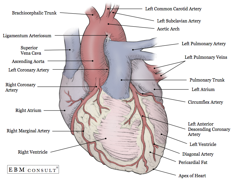
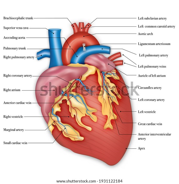
Heart: illustrated anatomy | e-Anatomy - IMAIOS This interactive atlas of human heart anatomy is based on medical illustrations and cadaver photography. The user can show or hide the anatomical labels which provide a useful tool to create illustrations perfectly adapted for teaching. ... Left ventricle, Left atrium, Anterior papillary muscle Figure 5 - Pericardium : Pericardial cavity ... Label the Heart Diagram Anterior view Quiz - PurposeGames.com Label the Heart Diagram Anterior view — Quiz Information. This is an online quiz called Label the Heart Diagram Anterior view. There is a printable worksheet available for download here so you can take the quiz with pen and paper. Anterior View Of Human Heart - Anatomy Note Anatomy note Odysee Channel, Please Subscribe to Support. Anterior View Of Human Heart In this image, you will find Brachiocephalic artery, left common carotid artery, left subclavian artery, aortic arch, superior vena cava, right pulmonary artery, ligamentum arteriosum, left pulmonary artery, left pulmonary veins in it. Heart anatomy: Structure, valves, coronary vessels | Kenhub Heart anatomy. The heart has five surfaces: base (posterior), diaphragmatic (inferior), sternocostal (anterior), and left and right pulmonary surfaces. It also has several margins: right, left, superior, and inferior: The right margin is the small section of the right atrium that extends between the superior and inferior vena cava .
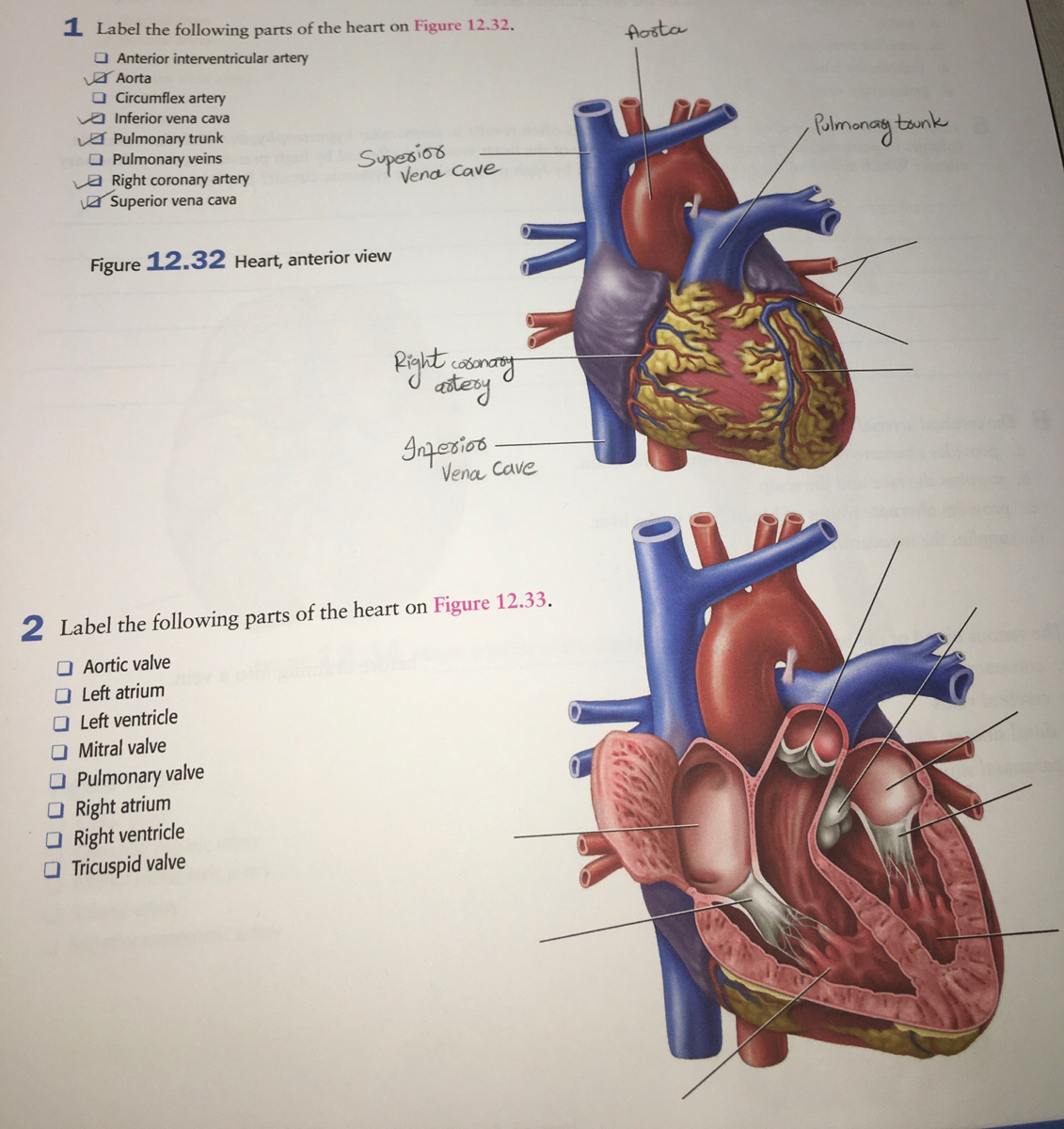
Answered: Label the following parts of the heart… | bartleby
anterior heart Quiz - PurposeGames.com Main Veins of the Human body. Science. Creator. goreydaydreams. Quiz Type. Image Quiz. Value. 22 points. Likes. 43. Played. ... This is an online quiz called anterior heart. ... quiz. heart. label. anterior. Quiz Points. 16 p. You need to get 100% to score the 16 points available. Game of the Day. Star Wars Chronology . by Geographonic. 945 plays.
Anatomy of the Human Heart - Physiopedia
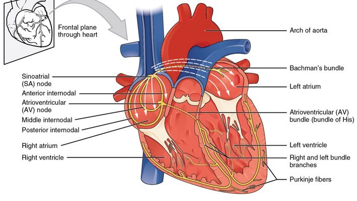
Chapter 22 Heart Flashcards | Quizlet Label the coronary arteries in an anterior view of the heart. Label the order that blood flows through in the heart, using the arrows as guides. Label the components of the heart wall. Label the components of the heart as seen from a posterior view. Label the major coronary veins. Label the components of the conduction system.
Sketch and label the ventral (anterior) view of human heart ...
Diagram of Human Heart and Blood Circulation in It Four Chambers of the Heart and Blood Circulation. The shape of the human heart is like an upside-down pear, weighing between 7-15 ounces, and is little larger than the size of the fist. It is located between the lungs, in the middle of the chest, behind and slightly to the left of the breast bone. The heart, one of the most significant organs ...
Untitled
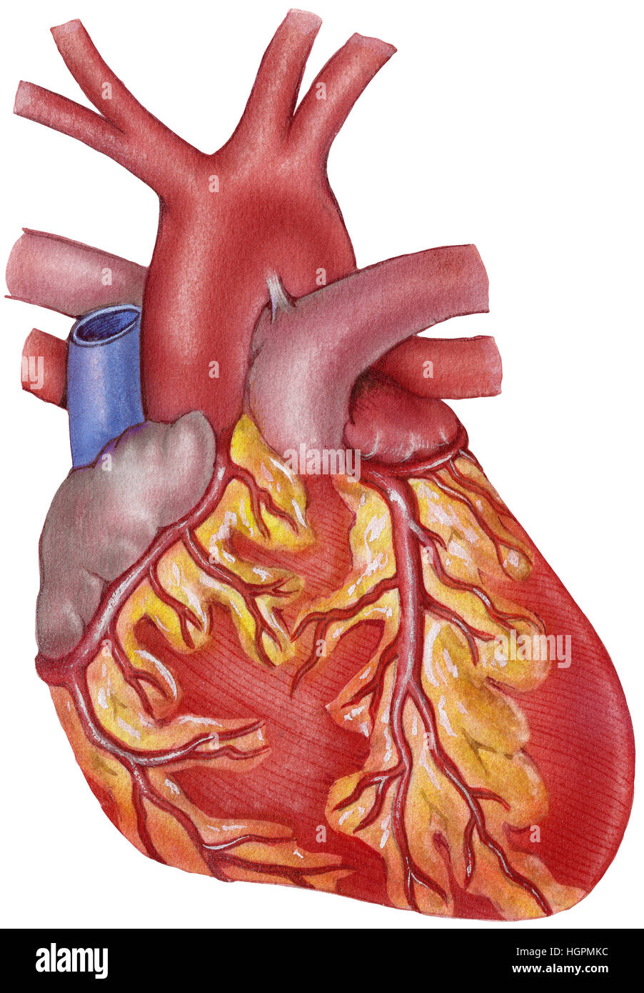
Anatomy: Heart (External) Orientation of the Heart Anterior or Sternocostal Surface: Mainly the right ventricle Inferior Border or Diaphragmatic Surface: Mainly the left ventricle and part of the right ventricle Right Border or Pulmonary Surface: Right atrium Left Border or Pulmonary Surface: Left ventricle and creates the cardiac impression in the left lung Blood Supply

Figure 35.2 Heart (dorsal view) Diagram | Quizlet
Lab Report 38 Figures 38.1, 38.2, and 38.3.pdf - Figure 38.1- Label ... Figure 38.3- Label this frontal section of the human heart. The arrows indicate the direction ofblood flow. 1.) Superior vena cava2.) Aorta3.) Right atrium4.) Aortic valve5.) Tricuspid valve 6.) Right ventricle7.) Interventricular septum 8.) Left atrium9.) Mitral valve10.) Left ventricle 11.)
Heart - Passnownow
Heart Lab Flashcards | Quizlet Label the features of the heart using the hints provided. Label the chambers and valves seen in an anterior view of the heart. Fill in the blanks with the appropriate words to describe blood flow from the heart. Then place the sentences in order to form a coherent paragraph. Label the coronary arteries on the posterior surface of the heart.
Heart anterior view hi-res stock photography and images - Alamy
Solved Label the structures seen in the anterior view of the - Chegg Question: Label the structures seen in the anterior view of the heart. Superior vena cava Interior vena cava Aorta Left atrium Pulmonary trunk Pulmonary vein Left ventricle Pulmonary artery Right ventricle Right atrium This problem has been solved! You'll get a detailed solution from a subject matter expert that helps you learn core concepts.

11 A&p ideas | anatomy and physiology, physiology, human ...
Gross anatomy of human heart anterior view We are pleased to provide you with the picture named Gross anatomy of human heart anterior view. We hope this picture Gross anatomy of human heart anterior view can help you study and research. for more anatomy content please follow us and visit our website: . Anatomynote.com found Gross anatomy of human heart anterior view ...
Anterior view of human heart anatomy with annotations Stock ...
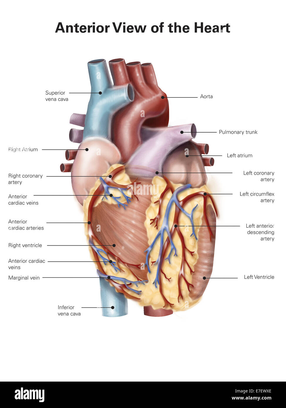
Anterior External View of Heart Labeling Diagram | Quizlet Anterior External View of Heart Labeling 5.0 (1 review) + − Learn Test Match Created by randajsmith Terms in this set (14) Superior Vena Cava ... Superior Vena Cava (SVC) ... Right Pulmonary Artery ... Right Pulmonary Veins ... Right Atrium ... Right Coronary Artery ... Right Ventricle ... Aorta ... Left pulmonary artery ... Left Atrium ...

Cardiovascular System | Human Anatomy | Life Science ...
Gross Anatomy of Heart - Diagram Anterior View Flashcards - Quizlet Gross Anatomy of Heart - Diagram Anterior View Flashcards Learn Test Match Flashcards Learn Test Match Created by bk2811 MVHS HAP HEART Terms in this set (26) Brachiocephalic trunk 1 Superior vena cava 2 Right pulmonary artery 3 Ascending aorta 4 Pulmonary trunk 5 Right pulmonary veins 6 Right atrium 7 Right coronary artery (in coronary sulcus) 8
Anterior view of the human heart Stock Photo - Alamy
Human Heart - Diagram and Anatomy of the Heart - Innerbody The AV valve on the right side of the heart is called the tricuspid valve because it is made of three cusps (flaps) that separate to allow blood to pass through and connect to block regurgitation of blood. The AV valve on the left side of the heart is called the mitral valve or the bicuspid valve because it has two cusps.
Solved g p rarte er interventi card wins middle and in ...

Anatomy of the Heart | Human heart anatomy, Gross anatomy ...

Heart Anatomy | Anatomy and Physiology II
Pre-lab 7 – Human Anatomy Lab Manual
Coronary Arteries | GetBodySmart

Heart Anatomy: Labeled Diagram, Structures, Blood Flow ...

anterior view of human heart Diagram | Quizlet
Anatomy: Heart (External)

Label the following diagram of the heart. | Homework.Study.com

File:Diagram of the human heart (cropped).svg - Wikimedia Commons

Anterior View of the Human Heart 35.1 Flashcards | Quizlet

Cardiology

Draw a diagram of the human heart and label its parts
Heart anterior view hi-res stock photography and images - Alamy
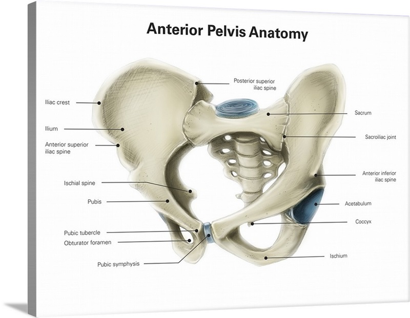
Anterior view of human pelvis, with labels Wall Art, Canvas ...

Human heart anatomy — atrium, Digitally Generated Image ...
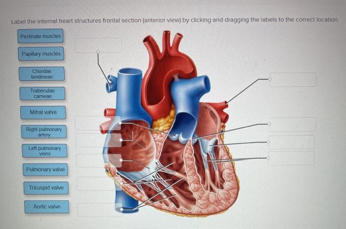
Solved Label the internal heart structures frontal section ...

Heart - Wikipedia
New Page 1
Sketch and label the dorsal (posterior) view of human heart ...
What is the difference between an aorta and a pulmonary ...
Heart Structure
Lesson | The Heart - External Structure | Encounter Edu
Heart Anatomy: Labeled Diagram, Structures, Blood Flow ...

Procedure A The Human Heart - Human Anatomy - GUWS Medical
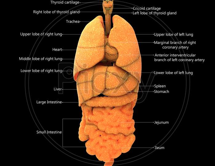
Image of Human Internal Organs Described with Labels Anatomy ...
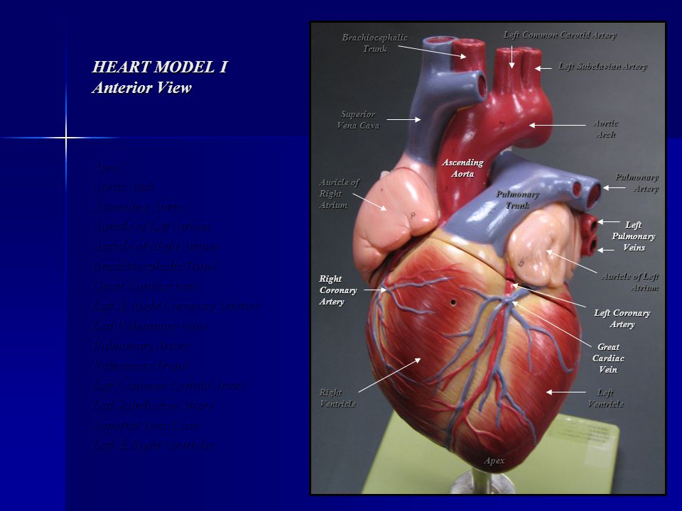
HEART MODEL I Anterior View
Heart diagram Images - Search Images on Everypixel
0 Response to "39 label this anterior view of the human heart"
Post a Comment