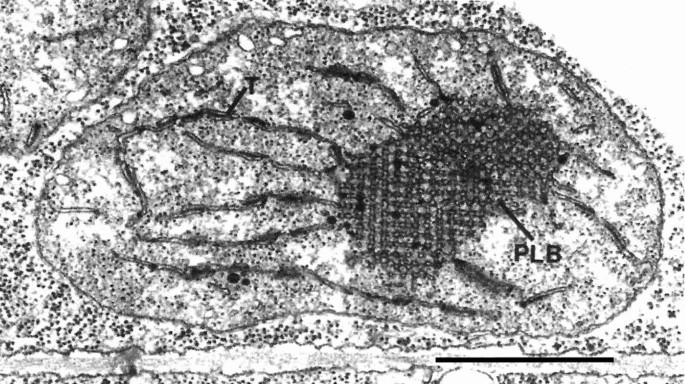
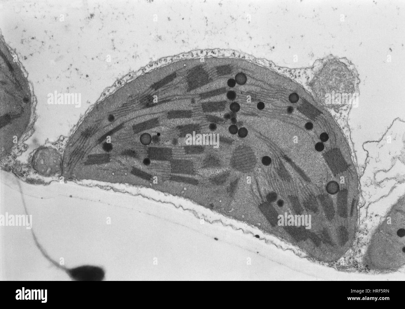
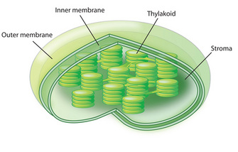
44 label the transmission electron microscope image of a chloroplast below
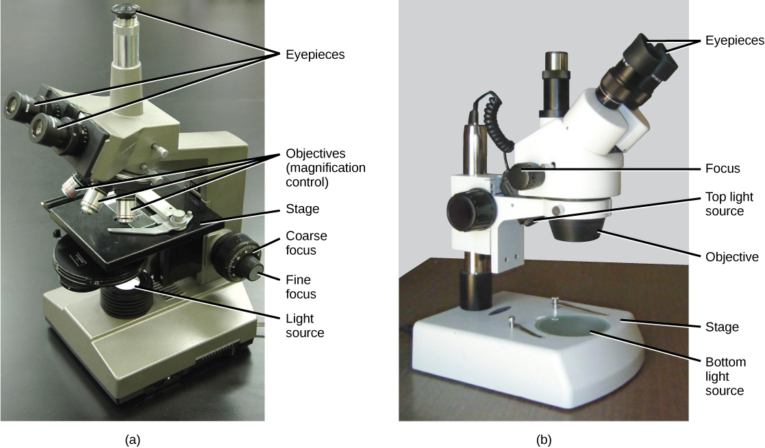
Scanning Electron Microscopic Study of Modified Chloroplasts in ... (TEM) to better understand the process of chloroplast degradation, ... Light micrographs of chloroplasts in the sepal cells of broccoli florets during ... Labeling A Microscope AnswersThis printable worksheet of ... The instructor will make note of incorrect and correct answers. microscope worksheet parts labeling science functions label pdf quiz worksheets labeled simple labels. The coarse adjustment knob is used to move the stage up and down. Supports the slide being viewed. com Label Microscope Diagram Using the terms listed below, label the microscope ...
Cells and magnification Flashcards | Quizlet c- A transmission electron microscope was used to produce the image in the figure ... 9) The figure below shows a photograph of a chloroplast taken with an ...

Label the transmission electron microscope image of a chloroplast below
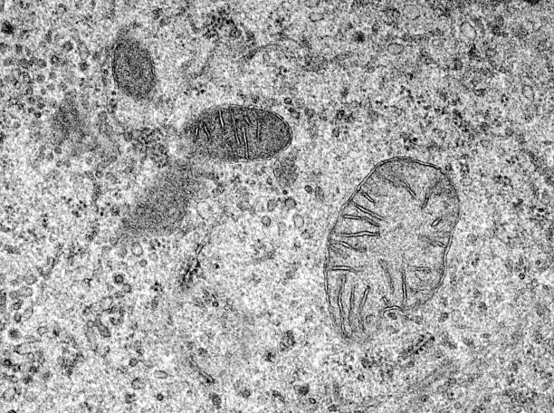
A-level Biology Bridging Course - Week 1 - Amazon S3 The images below are from a scanning electron microscope, transmission electron microscope and a ... (a) Name the parts of the chloroplast labelled A and B. Chloroplast hi-res stock photography and images - Alamy Find the perfect chloroplast stock photo, image, vector, illustration or 360 ... Transmission electron microscope image of a thin section cut from Begonia ... What are the labels of the transmission electronic microscope image ... Jul 28, 2021 ... What are the labels of the transmission electronic microscope image ... human mitochondria and the chloroplasts (plastids) and mitochondria.
Label the transmission electron microscope image of a chloroplast below. (PDF) Cambridge International AS and A Level Biology ... BIO1: Maintaining a Balance 1. Most organisms are active in a limited temperature range IDENTIFY THE ROLE OF ENZYMES IN METABOLISM, DESCRIBE THEIR CHEMICAL COMPOSITION AND USE A SIMPLE MODEL TO DESCRIBE THEIR SPECIFICITY ON SUBSTRATES 3.2 Cells // 3.2.1 Cell structure // 3.2.1.3 Methods of studying ... - Quizlet Image: Name the parts of the chloroplast labelled A and B ... The scientists used a transmission electron microscope to study the structure of the amoeba. Photosynthesis Equation. 6 CO 2 + 6 H 2 O + Light -> C 6 H 12 ... View photosynthesis_diagrams_worksheet from nursing psychiatri at our lady of fatima university. In the diagram below, label. Making energy chloroplasts photosynthesis is a. *Click On Open Button To Open And Print To Worksheet.Photosynthesis. 8.2.1 Draw and label a diagram showing the structure of a chloroplast as seen in electron micrographs. I ntroduction to Photosynthesis - images They account for 20% of the thylakoid membranes. 1. Based on the information above, label the transmission electron micrograph (TEM) of a chloroplast below: 2.
Top 14 Types of Spectroscopic Techniques – Explained ... ADVERTISEMENTS: Some of the important types of Spectroscopic Techniques are as follows: Type # 1. Gamma Spectroscopy: Gamma spectroscopy is a radionuclide measurement method. While a Geiger counter determines only the count rate, a gamma spectrometer will determine the energy and the count rate of gamma-rays emitted by radioactive substances. Gamma spectroscopy is an extremely […] Lifestyle | Daily Life | News | The Sydney Morning Herald The latest Lifestyle | Daily Life news, tips, opinion and advice from The Sydney Morning Herald covering life and relationships, beauty, fashion, health & wellbeing AP Biology Study the electron micrographs on page 110 of your text. ... Now label the diagram of the chloroplast below being to include the outer membrane, ... Transmission electron microscopic images of chloroplasts and... Ultrastructure of chloroplasts and mitochondria in wild-type cells. For easy organelle identification, a chloroplast (P) and a mitochondrion (M) are labeled. ( ...
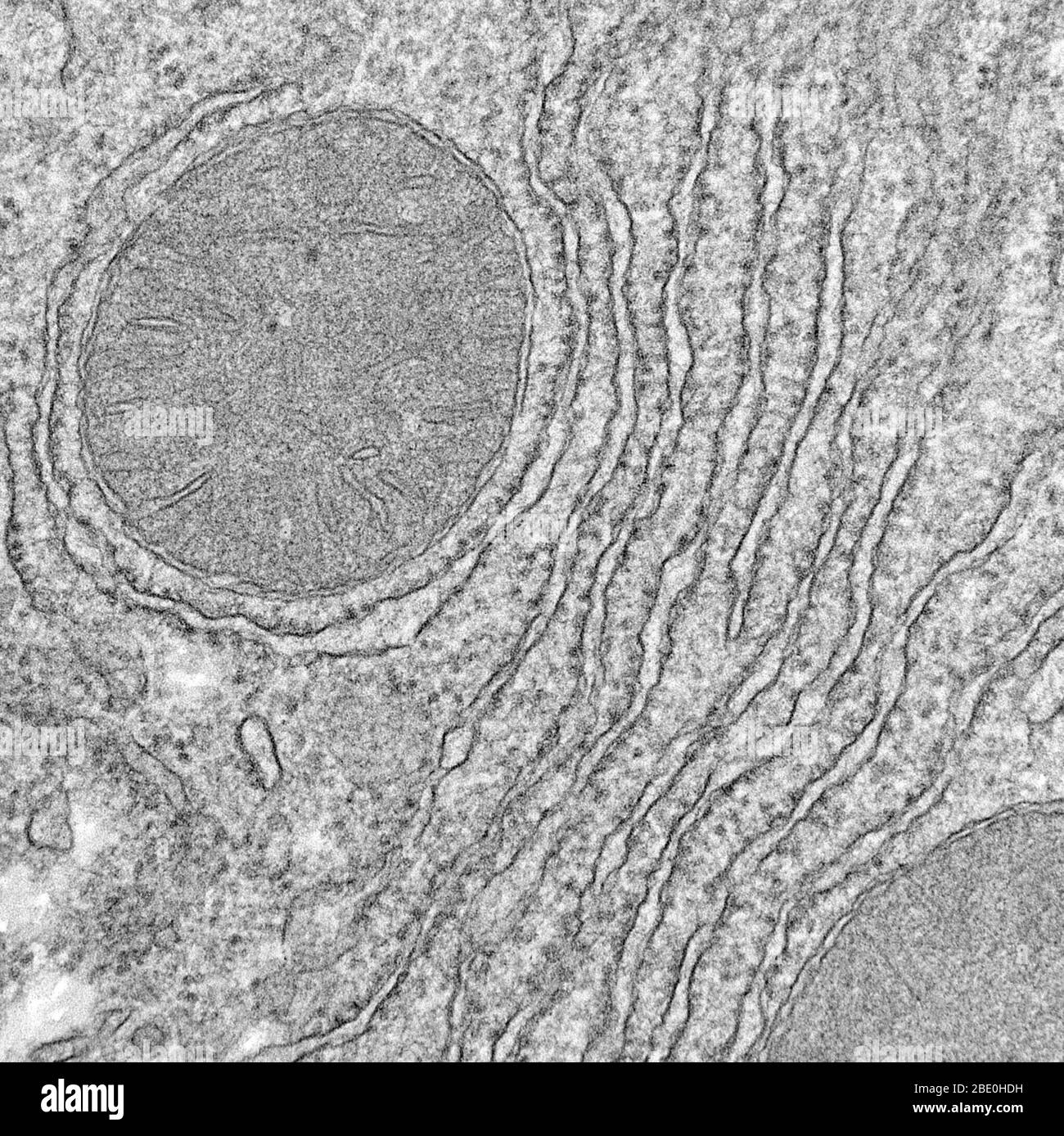
Bacillus subtilis biofilm matrix components target seed oil ... Jun 06, 2022 · The samples were left to dry and imaged under an FEI Tecnai G2 20 TWIN transmission electron microscope at an accelerating voltage of 80 kV. The images were acquired using TIA FEI Imaging Software ... Transmission electron microscopy picture of the chloroplast ... Transmission electron microscopy picture of the chloroplast envelopes. Highly magnified view of the outer area of a chloroplast displaying contact sites ... Nelson Biology 12.pdf [30j71j2z320w] - doku.pub The orbital that is occupied by an electron is what determines the energy level of the electron. The farther away the electron is from the nucleus, the greater its energy. The balloon-like 2p orbitals contain electrons that are farther away from the nucleus than the electrons in the 2s orbital, and thus hold the electrons with a higher energy ... What are the labels of the transmission electronic microscope image ... Jul 28, 2021 ... What are the labels of the transmission electronic microscope image ... human mitochondria and the chloroplasts (plastids) and mitochondria.
What are the labels of the transmission electronic microscope ...
Chloroplast hi-res stock photography and images - Alamy Find the perfect chloroplast stock photo, image, vector, illustration or 360 ... Transmission electron microscope image of a thin section cut from Begonia ...

DPS1 regulates cuticle development and leaf senescence in ...
A-level Biology Bridging Course - Week 1 - Amazon S3 The images below are from a scanning electron microscope, transmission electron microscope and a ... (a) Name the parts of the chloroplast labelled A and B.
A brief history of how microscopic studies led to the ...
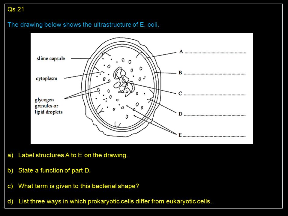
Qs 21 The drawing below shows the ultrastructure of E. coli.
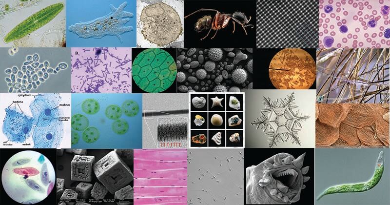
Amazing 27 Things Under The Microscope With Diagrams

Assignment 6, page 2

DP Biology: Ultrastructure of cells quiz 1.2
f i1w,-
A brief history of how microscopic studies led to the ...

Draw a labelled diagram of chloroplast as seen under an ...

1.2 Difference between plant and animal cells | Cells as the ...

Assignment 6, page 2

Cells and magnification Flashcards | Quizlet
3.1 How Cells Are Studied – Concepts of Biology – 1st ...
Electron micrograph chloroplast hi-res stock photography and ...
Tem mitochondria hi-res stock photography and images - Alamy
What is a diagram of a plant and animal cell under an ...

2.2 Molecular make up of cells | Cells: the basic units of ...

1.2 Difference between plant and animal cells | Cells as the ...
7,411 Electron Micrograph Stock Photos, Pictures & Royalty ...

Arabidopsis thaliana(L.) Accession Col | Science Lab | Leica ...
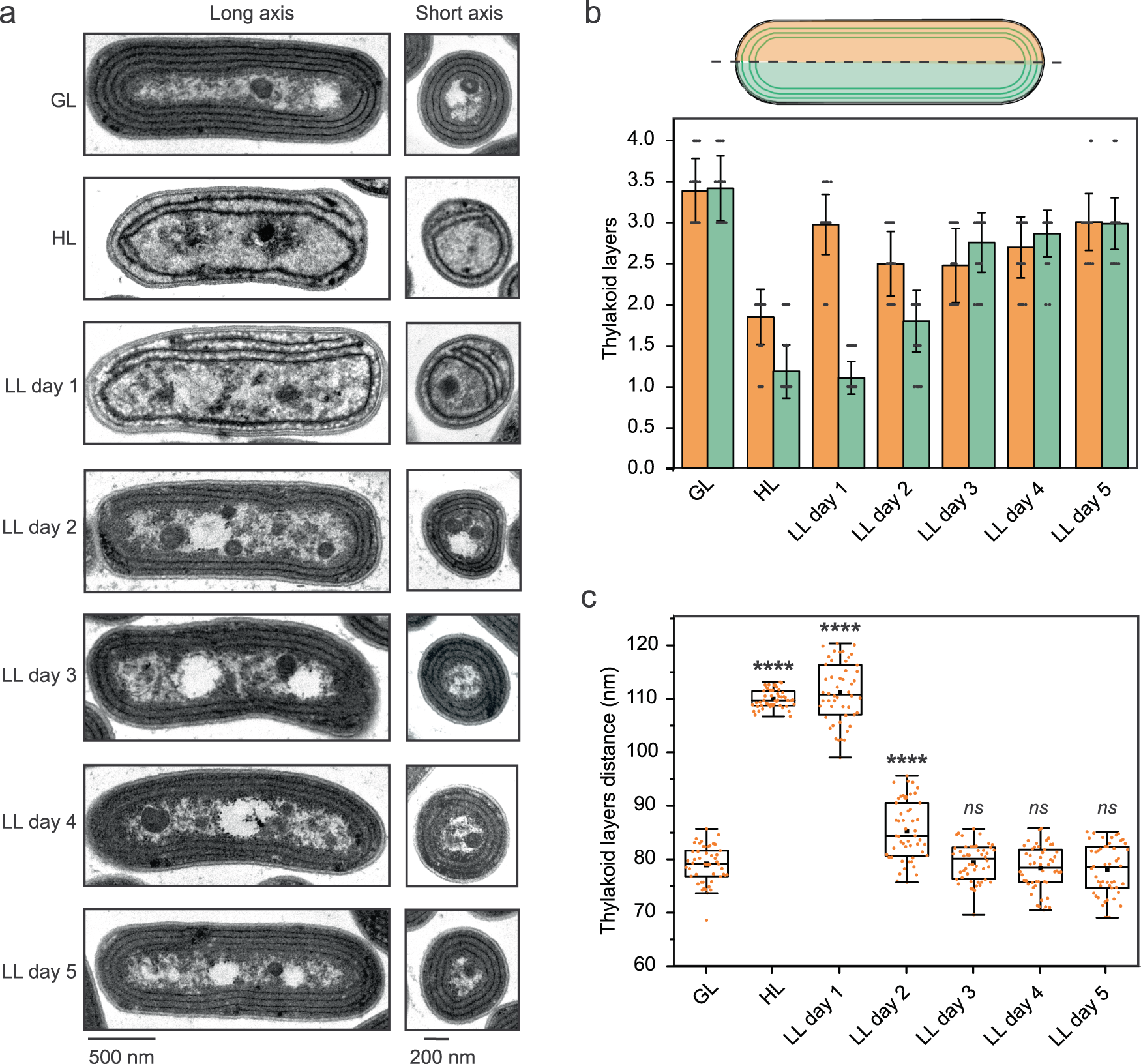
Probing the biogenesis pathway and dynamics of thylakoid ...

TEM vs SEM - Electron Microscopes - Advancing Materials
Plant cell | Definition, Characteristics, & Facts | Britannica

Endomembrane System | Biology for Majors I
Photosynthesis, Chloroplast | Learn Science at Scitable

microscopy how a microscope works magnification calculations ...

DP Biology: Ultrastructure of cells quiz 1.2

Arabidopsis thaliana(L.) Accession Col | Science Lab | Leica ...

Bio Photosynthesis: Chloroplast structure under a microscope ...
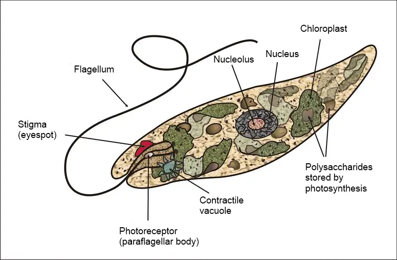
Euglena under the Microscope - Structure, Morphology ...
GR 9 Topic 7 Microscopy - AMAZING WORLD OF SCIENCE WITH MR. GREEN
f i1w,-

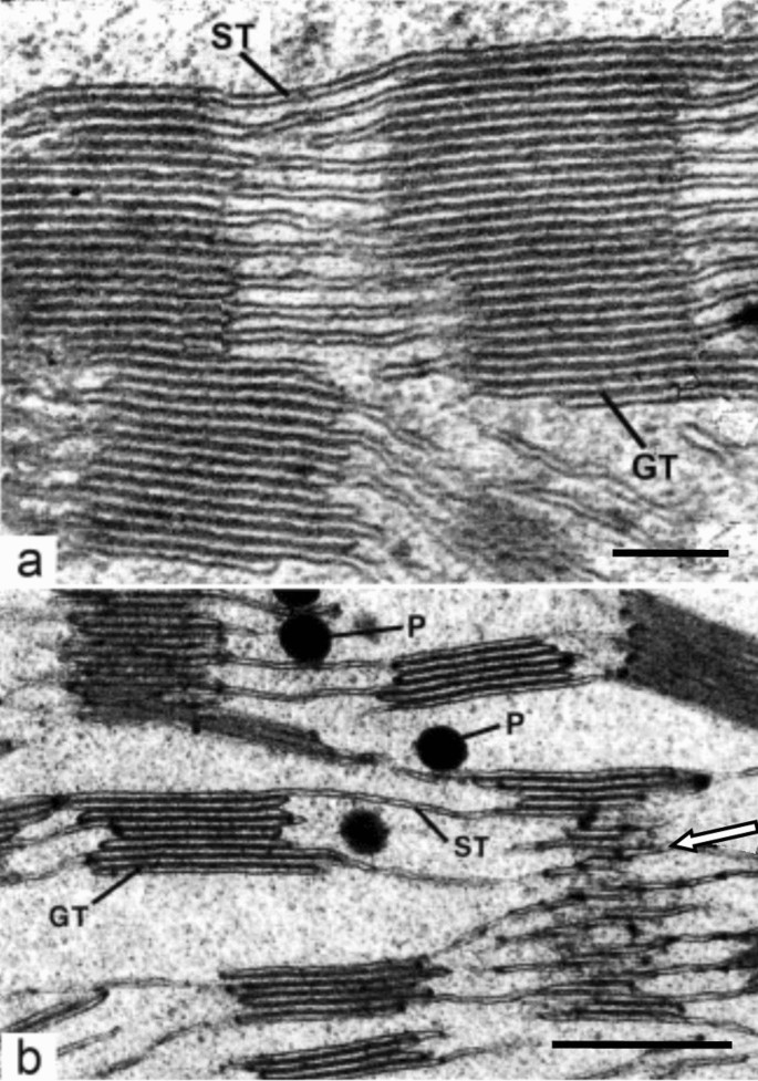
Electron micrograph of an Arabidopsis leaf chloroplast ...

Electron micrograph of an Arabidopsis leaf chloroplast ...
What is a diagram of a plant and animal cell under an ...

File:Lettuce Thylakoid.jpg - Wikipedia

Electron micrograph of isolated chloroplasts with the major ...

Label parts for diagrammes given below? And identify which ...

Draw a diagram of each of the following cells: a bacteria ...

Introduction to Photosynthesis

What Living Things You Can See Under a Light Microscope? - Rs ...
What is a diagram of a plant and animal cell under an ...

Cell Micrographs | BioNinja
0 Response to "44 label the transmission electron microscope image of a chloroplast below"
Post a Comment